Download
1 / 1
10 likes | 107 Views
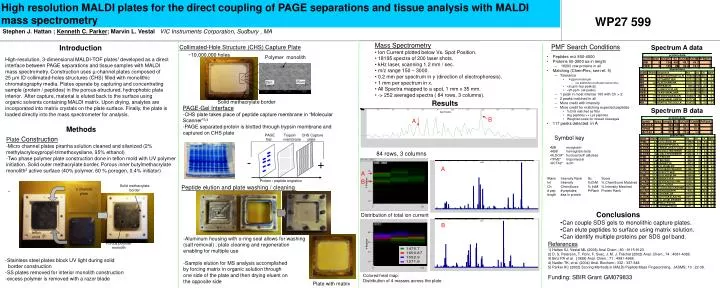
High resolution MALDI plates for the direct coupling of PAGE separations and tissue analysis with MALDI mass spectrometry. Polymer monolith. -. 35um. +. 25m. Solid methacrylate border. Solid methacrylate border. u channel plate. teflon mold. Porous polymer monolith. WP27 599.

E N D
High resolution MALDI plates for the direct coupling of PAGE separations and tissue analysis with MALDI mass spectrometry Polymer monolith - 35um + 25m Solid methacrylate border Solid methacrylate border u channel plate teflon mold Porous polymer monolith WP27 599 Stephen J. Hattan ; Kenneth C. Parker; Marvin L. VestalVIC Instruments Corporation, Sudbury , MA • Mass Spectrometry • Ion Current plotted below Vs. Spot Position. • 18195 spectra of 200 laser shots. • kHz laser, scanning 1.2 mm / sec. • m/z range 150 – 3000. • 0.2 mm per spectrum in y (direction of electrophoresis). • 1 mm per spectrum in x. • All Spectra mapped to a spot, 1 mm x 35 mm. • -> 252 averaged spectra ( 84 rows, 3 columns). PMF Search Conditions Spectrum A data Collimated-Hole Structure (CHS) Capture Plate Introduction ~10,000,000 holes High-resolution, 3-dimensional MALDI-TOF plates1 developed as a direct interface between PAGE separations and tissue samples with MALDI mass spectrometry. Construction uses µ-channel plates composed of 25 µm ID collimated-holes structures (CHS) filled with monolithic chromatography media. Plates operate by capturing and concentrating sample (protein / peptides) in the porous-structured, hydrophobic plate interior. After capture, material is eluted back to the surface using organic solvents containing MALDI matrix. Upon drying, analytes are incorporated into matrix crystals on the plate surface. Finally, the plate is loaded directly into the mass spectrometer for analysis. Results PAGE-Gel Interface -CHS plate takes place of peptide capture membrane in “Molecular Scanner”3,4 -PAGE separated protein is blotted through trypsin membrane and captured on CHS plate Spectrum B data B A in A Methods PAGE Trypsin CHS Capture Gel membrane plate Symbol key Plate Construction -Micro channel plates piranha solution cleaned and silanized (2% methylacryloxypropyl-trimethoxysilane, 95% ethanol) -Two phase polymer plate construction done in teflon mold with UV polymer initiation. Solid outer methacrylate border, Porous inner butylmethacrylate monolith2 active surface (40% polymer, 60 % porogen, 0.4% initiator) - • MB myoglobin • HBB hemoglobin beta • ALDOA* fructose bisP aldolase • TPM2* tropomyosin • ACTA2* actin 84 rows, 3 columns A A Protein / peptide migration B Peptide elution and plate washing / cleaning • Conclusions • Can couple SDS gels to monolithic capture plates. • Can elute peptides to surface using matrix solution. • Can identify multiple proteins per SDS gel band. Distribution of total ion current B • -Aluminum housing with o-ring seal allows for washing • (salt removal) , plate cleaning and regeneration • enabling for multiple use • Sample elution for MS analysis accomplished • by forcing matrix in organic solution through • one side of the plate and then drying eluent on • the opposite side References 1) Hattan SJ, Vestal ML (2008) Anal Chem.; 80 : 9115-9123. 2) D. S. Peterson, T. Rohr, F. Svec, J. M. J. Fréchet (2002) Anal. Chem.; 74 : 4081-4088. 3) Binz PA et al. (1999) Anal. Chem.; 71 : 4981-4988. 4) Nadler TK, et al. (2004) Anal. Biochem.; 332 : 337-348. 5) Parker KC (2002) Scoring Methods in MALDI Peptide Mass Fingerprinting.. JASMS; 13 : 22-39. Funding: SBIR Grant GM079833 -Stainless steel plates block UV light during solid border construction -SS plates removed for interior monolith construction -excess polymer is removed with a razor blade Colored heat map: Distribution of 4 masses across the plate Plate with matrix