Download
1 / 41
420 likes | 447 Views
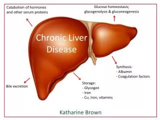
BIOCHEMICAL INVESTIGATIONS IN CHRONIC LIVER DISEASE. COL MUHAMMAD ASIF NAWAZ DEPARTMENT OF PATHOLOGY ARMY MEDICAL COLLEGE, RAWALPINDI. Normal Liver Function. Protein synthesis and degradation: albumin, transport proteins, clotting factors, Carbohydrate metabolism Lipid metabolism

E N D
BIOCHEMICAL INVESTIGATIONS IN CHRONIC LIVER DISEASE COL MUHAMMAD ASIF NAWAZ DEPARTMENT OF PATHOLOGY ARMY MEDICAL COLLEGE, RAWALPINDI
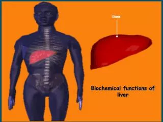
Normal Liver Function • Protein synthesis and degradation: • albumin, transport proteins, clotting factors, • Carbohydrate metabolism • Lipid metabolism • Bile acid metabolism • Bilirubin metabolism • Hormone inactivation • Drug inactivation and excretion • Storage function
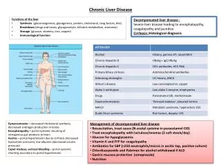
Types of liver disease • Cholestasis: bile duct damage from stones or tumour, primary biliary cirrhosis • Infection: hepatitis A, B, C, EBV, CMV • Chemical damage: drugs and alcohol • Hereditary: Wilsons disease, haemochromatosis • Vascular damage: Budd-Chiari • Autoimmunity: autoimmune hepatitis, primary sclerosing cholangitis • Congenital anomalies: biliary atresia, Caroli’s disease • Metabolic disease: galactosemia, fatty liver disease
Chronic Liver Disease • Chronic Viral hepatitis: B & C • Non-alcoholic fatty liver disease (NAFLD) • Alcohol • Autoimmune – autoimmmune hepatitis, PBC (Primary Biliary cirrhosis), PSC (Primary Sclerosing Cholangitis) • Haemochromatosis • Drugs (MTX, amiodarone) • Cystic fibrosis, a1antitryptin deficiency, Wilsons disease, • Vascular problems (Portal hypertension + liver disease) • Cryptogenic • Others: sarcoidosis, amyloid, schistosomiasis
Liver function tests • Noninvasive method of screening for the presence of liver dysfunction • Pattern of lab test abnormality allows recognition of general type of disorder • To assess the severity and occasionally allow prediction of outcome • To follow the course of the disease, evaluate response to treatment, and adjust treatment when necessary
Transaminases • Most sensitive indicator of liver injury • Participate in gluconeogenesis, transfer of amino groups from aspartate or alanine to ketoglutaric acid to form oxaloacetete or pyruvate. • AST present in cytosol and mitochondria in liver, cardiac muscle, skeletal muscle, kidney, brain, pancreas, lungs, WBC and RBC. AST is an early marker of liver damage • ALT a cytosolic enzyme, highest concentration in the liver, so liver specific but longer half life
Elevated ALT (SGPT) and AST (SGOT) levels • AST Mild elevations (<10 times UNL) • chronic viral hepatitis • nonalcoholic steatohepatitis • fatty liver • liver cirrhosis • Moderate and marked elevations(>10 times UNL) • acute viral hepatitis • Alcoholic liver disease • autoimmune hepatitis • toxic and drug-induced liver necrosis • Shock or ischemia to liver • AST/ALT ratio • < 1 in most hepatocellular injury • >1 in alcholic liver diseae, drug induced, malignancy, cirrohosis
Enzymes for the detection of cholestasisAlkaline phosphatase • Present in nearly all tissues - isoenzymes • Localised in the microvilli of the bile canalicus in the liver • Also present in bone, intestine, placenta, kidney and wbc • Elevation may be physiological or pathological • Physiological • In tissues undergoing metabolic stimulation • Third trimester of pregnancy • Adolescence
Suggested algorithm for evaluating a raised s.alkaline phosphatase
Gammaglutamyl transferase (γ-glutamyl transpeptidase) • Found in hepatocytes and biliary epithelial cells • Sensitive for hepatobiliary disease but ltd by lack of specificity • With other enzyme abnormalities, raised GGT would support a hepatobiliary cause • Can confirm hepatic source for a raised AP • Raised GGT and raised transaminases with ratio of AST to ALT 2:1 or more suggestive of ALD • Medications can cause mild rise • Normal range 0 to 30 IU/L
5´-Nucleotidase • Normal 0.3 to 3.2 Bodansky units • Spectrum of abnormality similar to that of SAP • Specificity for hepatobiliary disease • May be used to confirm hepatic origin of elevated SAP
Lactate Dehydrogenase • Lactate dehydrogenase (LDH) is often raised in hepatocellular dysfunction • It is rarely measured for this purpose since it lacks specificity because of wide distribution of LDH in the body.
Serum Bilirubin • A breakdown product of heme (part of the haemoglobin in red blood cells) • The hepatocytes takes up bilirubin, conjugates it to make it more water soluble and secretes it onto the bile ducts for excretion via the intestine • Increased bilirubin causes jaundice • Prehepatic: too much red cell break down (unconjugated) • Hepatic: unable to metabolise the bilirubin (mixed) • Reduced conjugation • Unable to secrete bilirubin • Post hepatic: obstruction to the excretion of bile (conjugated)
Causes of bilirubin elevation • Liver disease: usually along with other LFTs • Isolated ↑ bilirubin: familial hyperbilirubinaemias, • Haemolysis: ↑ unconjugated bilirubin.
Follow up of elevated bilirubin levels (when no clinical indications of cause)
Liver synthetic functions • Clotting factors: prothrombin (PT) – very specific for liver dysfunction / liver failure • Albumin: low in chronic liver disease • Glucose: hypoglycaemia indicates severe hepatic dysfunction
Serum albumin • Synthesized by hepatocytes • Serum half life about 3 weeks • Decreased in chronic and severe liver disease • Other causes for hypoalbinemia: • protein-losing enteropathy • Urinary losses: nephrotic syndrome • malnutrition
Plasma Proteins in Liver Disease • Serum globulins are often increased in cirrhosis • alpha1-Antitrypsin deficiency: - neonatal jaundice and - cirrhosis in children and young adults.
Plasma Proteins in Liver Disease • Alpha-fetoprotein: - modest levels are found, e.g; during acute viral hepatitis, - very high values occur in hepatocellular carcinoma.
Autoantibodies • Antimitochondrial Ab: in primary biliary cirrhosis • Antinuclear Ab and antismooth muscle Ab: in autoimmune hepatitis type 1 • Anti LKM1 antibodies in type 2 AIH • Antibodies to soluble liver antigen in type 3 AIH
Serum ammonia • Released from proteins in the gut • Detoxified in the liver to urea • Increased serum level due to decreased detoxification by the liver and due to portal-systemic shunting • Elevation does not correlate with hepatic function or the presence or degree of hepatic encephalopathy.
Tumor markers • fetoprotein: increased in hepatocellular carcinoma. • CA 19-9: increased in tumors of biliary tree
Blood sugar in liver diseases • Impaired Glucose tolerance test in liver cirrhosis • Hypoglycemia in fulminant hepatitis and terminal liver cirrhosis
Serum lipids in liver disease • Cholesterol level increased in liver diseases especially cholestatic diseases with decreased esterified fraction. • Abnormal lipoprotein X in biliary cirrhosis. • Triglyceride level increased due to decreased mobilization from liver cells
Liver function tests 2 • Hepatitis antibodies: A, B, C….D, E • EBV, Toxo, CMV, Leptospirosis • Ferritin and fasting transferrin saturation, • Haemochromatosis genetics • Caeruloplasmin and copper (serum), • 24 hour urine for copper • Autoantibodies: ANA, ASMA, AMA, Coeliac • Immunoglobulins: IgG, IgA, IgM • Cholesterol, triglycerides, glucose, TFTs • a1antitrypsin levels + phenotype • a-fetoprotein (cirrhotics only)
Chronic hepatitis B • 50 - 90% neonates and children infected with hepatitis will develop chronic hepatitis B infection, but < 5% of adults. • Chronic hepatitis B carriers have ~ 25% risk of developing liver damage, cirrhosis, liver failure and liver cancer. • LFTs should be tested at least 6 monthly. • Screening for hepatitis B infection, using HBsAg, is recommended for all people not previously been immunised.
HBsAg Negative Positive No evidence of Hepatitis B infection If positive for > 6 months, consistent with chronic Hepatitis B infection Screening for hepatitis B in people not previously immune
Anti-HBs Negative Positive No evidence of previous infection or immunization See footnote(b) Compatible with previous infection or immunisation Investigation for Previous Infection or Immunisation with Hepatitis B – See footnote (a) • A previous vaccination with documented immune response, the patient can then be presumed to be protected long term unless they are immunosuppressed. If in doubt revaccinate and recheck anti-HBs in 3 weeks. • A small number of patients may be positive for anti-HBc from a previous HBV infection in the absence of anti-HBs. If there is a strong suspicion of previous infection or high risk, then order an anti-HBc.
Chronic hepatitis C- Most people will not be symptomatic during the acute infection but approximately 70% will remain infected. - Chronic infections carry a substantial risk of liver damage, cirrhosis and liver cancer.- Test blood for Anti HCV-Ab of all those at risk e.g:blood /components reciepients.
Anti-HCV Ab Negative Positive No evidence of chronic Hepatitis C infection See footnote (a) Indicates possible current, previous or chronic Hepatitis C infection See footnote (b) Investigation for Evidence of Chronic HCV Infection • A negative test does not exclude infection within the previous eight weeks. • Positive anti-HCV is followed up by HCV RNA tests. Persistently normal LFTs and two negative HCV RNA tests 3 months apart indicate that active HCV is extremely unlikely.
LFTs Requests Liver function testing is not indicated for asymptomatic people without risk factors
Asymptomatic people at risk of abnormal LFT’s • Diabetes or metabolic syndrome (increased risk of NAFLD) • Chronic hepatitis B • Chronic hepatitis C • Excessive alcohol intake
Risk of abnormal LFTs using drugs Drugs for which LFT monitoring is recommended in primary care:
Monitoring of LFTs for statin use • Risk of liver damage from statin use has been overstated. • Liver failure occurs with statins is similar to liver failure rate in general population. • Irreversible liver damage resulting from statin therapy is exceedingly rare. • Routine monitoring is not necessary. • Statins should not be withheld in patients with baseline abnormal LFTs.
Laboratory Findings in Progression of Chronic Hepatitis to Cirrhosis
Cirrhosis • MELD = 6.43 + 9.57 (Creatinine mg/dL max. upto 4 + 3.78 (Bilirubin mg/dL) + 11.2 (INR) • Score > 15 , liver transplantation may be considered