Download
1 / 46
940 likes | 2.16k Views
DNA-Based Tissue Typing. Chapter 15. Major Histocompatibility Complex. Major Histocompatibility Complex Cluster of genes found in all mammals Its products play role in discriminating self/non-self Participant in both humoral and cell-mediated immunity

E N D
DNA-Based Tissue Typing Chapter 15
Major Histocompatibility Complex • Major Histocompatibility Complex • Cluster of genes found in all mammals • Its products play role in discriminating self/non-self • Participant in both humoral and cell-mediated immunity • MHC Act As Antigen Presenting Structures • In humans MHC is found on chromosome 6 • Referred to as HLA complex • In mice, MHC is found on Chromosome 17 • Referred to as H-2 complex
The Major Histocompatibility Complex (MHC) • The MHC is located on chromosome 6. • The MHC contains the human leukocyte antigen (HLA) and other genes. 1 Mb 2 Mb 3 Mb 4 Mb TNF b a b a b a b b b b a a b HLA- DP DQ DR B C A Class II Class III Class I
Classes of MHC Genes • Class I MHC genes • Glycoproteins expressed on all nucleated cells • Major function to present processed Ags to TC • Class II MHC genes • Glycoproteins expressed on M, B-cells, DCs • Major function to present processed Ags to TH • Class III MHC genes • Products that include secreted proteins that have immune functions. Ex. Complement system, inflammatory molecules
Class I MHC Genes found in regions A, B and C in humans (K and D in mice) • Class II MHC Genes found in regions DR, DP and DQ (IA and IE In mice) • Class I and Class II MHC share structural features • Both involved in APC • Class III MHC have no structural similarity to Class I and II • Ex. TNF, heat shock proteins, complement components
MHC Genes Are Polymorphic • MHC products are highly polymorphic • Vary considerably from person to person • However, crossover rate is low • 0.5% crossover rate • Inherited as 2 sets (one from father, one from mother) • Haplotype refers to set from mother or father • MHC alleles are co-dominantly expressed • Both mother and father alleles are expressed
Class I MHC Molecule • Comprised of 2 molecules • chain (45 kDa), transmembrane • 2-microglobulin (12 kDa) • Non-covalently associated with each oth • Association of chain and 2 is required for surface expression • chain made up of 3 domains (1, 2 and 3) • 2-microglobulin similar to 3 • 1 and 2 form peptide binding cleft • Fits peptide of about 8-10 a/a long • 3 highly conserved among MHC I molecules • Interacts with CD8 (TC) molecule
Class II MHC Molecule • Comprised of and chains • chain and chain associate non-covalently • and chains made up of domains • 1 and 2 ( chain) • 1 and 2 ( chain) • 1and 1 form antigen binding cleft • and heterodimer has been shown to dimerize • CD4 molecule binds 2/2 domains
Class I And II Specificity • Several hundred allelic variants have been identified • However, up to 6 MHC I and 12 MHC II Molecules are expressed in an individual • Enormous number of peptides needs to be presented using these MHC molecules • To achieve this task MHC molecules are not very specific for peptides (unlike TCR and BCR) • Promiscuous binding occurs • A peptide can bind a number of MHC • An MHC molecule can bind numerous peptides
Class I And II Diversity And Polymorphism • MHC is one of the most polymorphic complexes known • Alleles can differ up to 20 a/a • Class I Alleles: 240 A, 470 B, 110 C • Class II Alleles: HLA-DR 350 , 2 ! • HLA-DR • genes vary from 2-9 in different individuals!!!, • 1 gene ( can combine with all products increasing number of APC molecules) • DP (2 , 2 ) and DQ (2 , 3 )
Class I MHC Peptides • Peptides presented thru MHC I are endogenous proteins • As few as 100 Peptide/MHC complexes can activate TC • Peptide Features • size 8-10 a/a, preferably 9 • Peptides bind MHC due to presence of specific a/a found at the ends of peptide. • Ex. Glycine @ position 2
Class II MHC Peptides • Peptides presented thru MHC II are exogenous • Processed thru endocytic pathway • Peptides are presented to TH • Peptides are 13-18 a/a long • Binding is due to central 13 a/a • Longer peptides can still bind MHC II • Like a long hot dog • MHC I peptides fit exactly, not the case with MHC II peptides
MHC Expression • Expression is regulated by many cytokines • IFN, IFN, IFN and TNF increase MHC expression • Transcription factors that increase MHC gene expression • CIITA (transactivator), RFX (transactivator) • Some viruses decrease MHC expression • CMV, HBV, Ad12 • Reduction of MHC may allow for immune system evasion
a b a a 1 1 2 1 HLA-D a b 2 2 a 3 b 2 microglobulin Cell membrane a b a chain chain chain Human Leukocyte Antigens (HLA) • Human leukocyte antigens, the MHC gene products, are membrane proteins that are responsible for rejection of transplanted organs and tissues.
Human Leukocyte Antigens (HLA) • HLA-gene sequences differ from one individual to another. • Also written as: • Each sequence is a different allele.
Subregion Gene region HLA-DRB1 a-or b-chain polypeptide Gene locus HLA Allele Nomenclature • A standard nomenclature has been established by the World Health Organization (WHO) Nomenclature Committee. • A small “w” is included in HLA-C, HLAB-4, and HLAB-6 allele nomenclature: HLA-Cw, HLABw-4, HLABw-6.
HLA Typing • Every person (except identical twins) has different sets of HLA alleles. • Transplanted organs are allografts, in which the donor organ and the recipient are genetically different. • Compatibility (matching) of the HLA of the donor and the recipient increases the chance for a successful engraftment. • Matching is determined by comparing alleles. • Resolution is the level of detail with which an allele is determined.
Lymphocytes from organ donor or lymphocytes of known HLA types Positive reaction to antibody kills cells: dead cells pick up dye. Recipient serum Negative reaction to antibody: cells survive and exclude dye. Serological Typing • Recipient antihuman antibodies are assessed by crossmatching to donor lymphocytes.
Serological Typing Using Bead Arrays • Recipient antihuman antibodies are assessed by crossmatching to known lymphocyte antigens conjugated to microparticles. Results are assessed by flow cytometry. Serum antibodies Beads conjugated to different lymphocyte antigens Positive for antibody (Wash) Fluorescent reporter antibodies Negative for antibody
Other Serological Typing Methods • Cytotoxic and noncytotoxic methods with flow cytometry detection. • Enzyme-linked immunosorbent assay (ELISA) with solubilized HLA antigens. • Mixed lymphocyte culture measuring growth of lymphocytes activated by cross-reactivity. • Measure of HLA-protein mobility differences in one-dimensional gel isoelectric focusing or two-dimensional gel electrophoresis.
ELISA • Class I and class II are solid phase enzyme linked immuno sorbent assays (ELISA). Microtitre plates are coated with different highly purified human HLA class I and II glycoproteins. If the sample being tested contains specific antibodies against HLA class I or class II, they will bind to the antigens in the wells of the microtitre plate.
ELISA • The resulting antibody-antigen complex is detected using a specific enzyme-labelled (alkaline phosphatase) antibody which is directed against human IgG (conjugate). The presence of bound antibodies is demonstrated by adding a chromogenic substrate (PNPP) which results in a coloured product. The reaction is interpreted by a photometric reader.
MLC • Measure of histocompatibility at the hl-a locus. Peripheral blood lymphocytes from two individuals are mixed together in tissue culture for several days. Lymphocytes from incompatible individuals will stimulate each other to proliferate significantly (measured by tritiated thymidine uptake) whereas those from compatible individuals will not.
MLC • In the one-way MLC test, the lymphocytes from one of the individuals are inactivated (usually by treatment with mitomycin c or radiation) thereby allowing only the untreated remaining population of cells to proliferate in response to foreign histocompatibility antigens.
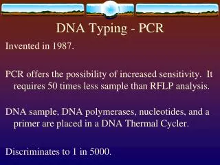
DNA-Based Typing Methods • DNA typing focuses on the most polymorphic loci in the MHC, HLA-B, and HLA-DRB. • Whole-blood patient specimens collected in anticoagulant are used for DNA typing. • Cell lines of known HLA type are used for reference samples.
Specimen 1 (Type A*0203) Specimen 2 (Type A*0501) TAG C GAT TAG A GAT ATC G CTA ATC T CTA Amplify, denature, and spot onto membranes Specimen 1 Specimen 2 Probe with allele-specific probes ...TAGCGAT..(A*02) ...TAGAGAT…(A*05) Specimen 2 Specimen 2 Specimen 1 Specimen 1 DNA-Based Typing Methods: SSOP • Sequence-specific oligonucleotide probe hybridization (SSOP, SSOPH)
PCR-SSO • Reverse SSO hybrodization is used to determine HLA-A, -B, -C, -DR, -DQ and -DP locus types at an intermediate level of resolution, somewhat higher than serological testing. Tests of this type are used when low or intermediate resolution typing is required or as a screening test to identify potential donors or individuals who may later require higher resolution testing. • This technology is used for high volume testing and allows for relatively low-cost typing for bone marrow donor drives or other applications involving large sample numbers. The laboratory can process as many as 25,000 samples per drive. Special volume pricing and terms may apply.
DNA-Based Typing Methods: SSP-PCR • Sequence-specific PCR is performed with allele-specific primers. Amplification controls SSP= Sequence-specific primer Allele-specific product SSP matches allele Amplification SSP No amplification SSP SSP does not match allele
PCR-SSP • PCR-SSP is also used to determine HLA-A, -B, -C, -DR and DQ locus types at a resolution similar to serological testing. PCR-SSP is a very rapid test that can be performed in 3-4 hours from the time a sample is received. PCR-SSP is used for typing deceased organ donors when speed is an important consideration. PCR-SSP can also be used to provide higher resolution testing and may be employed to resolve alleles.
Amplification control Allele-specific product Agarose gel DNA-Based Typing Methods: SSP-PCR • Primers recognizing different alleles are supplied in a 96-well plate format. Reagent blank
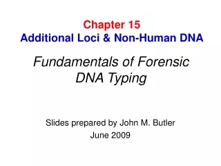
Reverse PCR primer Forward PCR primer Sequencing primers HLA-B DNA-Based Typing Methods: Sequence-Based Typing • Sequence-based typing (SBT) is high resolution. • Polymorphic regions are amplified by PCR and then sequenced.
SBT • SBT provides the highest resolution HLA typing for HLA-A, -B, -C, -DR, -DQ and -DP locus alleles. SBT is used when the highest resolution typing is important as in donors and recipients of stem cell transplants or in examining disease associations
Sequence-Based Typing Sequences are compared to reference sequences for previously assigned alleles.
Typing Discrepancies • DNA sequence changes do not always affect epitopes. • Serology does not recognize every allele detectable by DNA. • New antigens recognized by serology may be assigned to a previously identified parent allele by SBT. • Serology antibodies may be cross-reactive for multiple alleles. • Due to new allele discovery, retyping results may differ from typing performed before the new allele was known.
Combining Typing Results • SSP-PCR followed by PCR RFLP • SSOP followed by SSP-PCR • SBT results clarified by serology