Download
1 / 75
900 likes | 1.45k Views
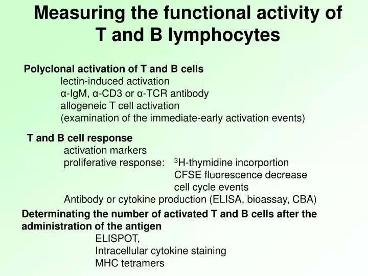
Measuring the functional activity of T and B lymphocytes. Polyclonal activation of T and B cells lectin-induced activation α-IgM, α-CD3 or α-TCR antibody allogeneic T cell activation (examination of the immediate-early activation events). T and B cell response activation markers

E N D
Measuring the functional activity of T and Blymphocytes Polyclonal activation of T and Bcells lectin-induced activation α-IgM, α-CD3 or α-TCR antibody allogeneicTcell activation (examination of the immediate-early activation events) T and Bcell response activation markers proliferative response: 3H-thymidine incorportion CFSE fluorescence decrease cell cycle events Antibody or cytokine production (ELISA, bioassay, CBA) Determinating the number of activated T and B cells after the administration of the antigen ELISPOT, Intracellular cytokine staining MHC tetramers
(review) Phases of the humoral immune response
(review) Phases of T cell response
Immunodeficiencies mainly characterized by different functional immunoassays Lymphocyte activation by specific antigen is hardly detected, because of the low number of the antigen specific cells Lymphocyte function can be investigated by polyclonal T/B-lymphocyte activator materials
Polyclonal activation of lymphocytes by LPS, lectins, PMA/ionomycin T cell C T cell B A B cell TLR4 (PMA activates protein kinase C) BCR or TCR-specifc antibodies could activate the lymphocytes also
Polyclonal B cell activators Activator T cell dependency Ig secretion Human B cells PWM (pokeweed mitogen) no yes SpA (superantigen, staphylococcus protein A) no yes EBV (transforming effect) yes yes Anti-Ig yes In the presence of cytokines
Pokeweed (PWM) (Phytolacca americana) – formerly used for coloring red wine (toxic: triterpene saponin) Chenopodiales Phytolaccaceae
Phytohaemagglutinin (PHA) Canavalia ensiformis – Jackbean, Sword bean
EXAMINATION OF T AND B CELL FUNCTIONS
Receptor crosslinking (immediate) phosphorylation steps (seconds-minutes) - Western blot - Bead array Antigen receptors (TCR, BCR), and different other receptors (eg. cytokine receptors) I.c Ca2+ increase - FACS, microscopy Gene activation - RT-PCR Cytokine synthesis - i.c cytometry • ELISA, ELISPOT • Bioassay Cytokine secretion - DNA content - IN antigens Cell-cycle/apoptosis Lymphocyte activation Cell division - 3H-thymidine, CFSE, MTT The examination often requires specific Ag-Ab reactions
Western blotting • Steps: • sample preparation (cells, tissues) • gel electrophoresis • blotting • labeling • development Anode(+) use: Identification of defined components from protein mixtures by antigen specific antibodies Cathode(-)
Western Blot • SDS-PAGE gel resolved into single protein bands (overlap possible) • Presence of a protein is determined by hybridizing the proteins, transferred or • applied to a membrane, with the relevant antibody Protein sample Standard Antibody recognizes epitope in specific protein Western blot Membrane SDS-PAGE
Western Blot • Used to detect specific proteins in a sample • Proteins separated by Sodium Dodecyl Sulfate-Polyacrylamide Gel Electrophoresis (SDS-PAGE), transferred to a membrane • Primary (1st) antibody (monoclonal or polyclonal) used to detect protein • Enzyme linked 2nd antibody (e.g. horseradish peroxidase-linked) used to detect 1st antibody
Investigation of the presence or absence of Bruton’s tyrosine kinase (BTK) by Western blot X-linked agammaglobulinemia. XLA patients do not generate mature B cells, which manifests as an almost complete lack of antibodies in their bloodstream.
Investigation of the presence or absence of Bruton’s tyrosine kinase (BTK) by flow cytometry Futatani T et al. Blood 1998;91:595-602
Detection of intracellular Ca2+ concentration An increase in cytoplasmic Ca2+ levels can be detected by fluorescent indicator dyes. /Fluo-3 or Indo-1/ Fluo-3 AM – excitable by blue light Indo-1 AM – excitable by UV light These indicator dyes bound to apolar groups (e.g. acetoxy-methylester: AM)cross the cell membrane,in the cell, esterases cleve them so the fluorochromesbecome polar andare trapped in the cell
e.g. Fluo-3 or Indo-1 Fluorescence proportional with Intracellular Ca2+level activation of cells time basic signal Measurement of Ca2+ signal by flow cytometry You can detect by fluorimeter also
Measurement of Ca2+ signal by flow cytometry T cell hybridoma specific for influenza virus hemagglutinin protein-derived peptide - Ca2+signal by antigen presentation activated T cells T cell activation (APC - T cell) non-activated T cells
Immunohistochemistry Labeled antibodies added to fixed tissue sections detect the distribution of the chosen antigen within the tissue or within the cells of a particular tissue • Immunofluorescence • Fluorescent dye coupled to antibody • FITC – fluorescein isothiocyanate (green) • PE – phycoerythrin (orange) • Immunoenzyme method • enzyme-coupled antibody • P – peroxidase • PA – alkaline phosphatase • (Substrates converted into an insoluble compound)
Immunohistochemistry Fixation Sectioning Tissue sample Section before staining Freezing
ImmunohistochemistryABC Method Enzim Avidin X Biotin Secondary antibody Primary antibody Slide Cells Tissue sample
Classical histochemistry Acute bronchopneumonia (hematoxilin- eosin staining)
Immunohistochemistry (CD68+ macrophages and lymphocytes, granuloma)
Antinuclear autoantiboies(ANA)from the serum of a SLE patient can be visualized in cell culture (Hep-2) by indirect fluorescent labeling (immunofluorescence)
Immunohistochemistry using fluorescent detection Detection of actin microfilaments
A fixed and permeabilized skin fibroblast. Mitochondriawere labeled with mouse IgG (anti–OxPhos Complex V) and visualized using goat anti–mouse IgG conjugated with orange-fluorescent Alexa Fluor 555. F-actinwaslabeled with green-fluorescent Alexa Fluor 488 phalloidin (a mushroom toxin). Nucleuswas stained with TO-PRO-3 iodide.
Peroxisome labeling in fixed and permeabilized pulmonary arteryendothelial cell.Peroxisomeswere labeled using an antibody directed at peroxisomal membrane protein 70 and detected with Alexa Fluor 488–labeled goat anti–mouse IgG. Mitochondria were stained with MitoTracker Red prior to fixation; Nuclei were stained with blue-fluorescent DAPI.
Flow cytometry An immunofluorescent method that mutually complements the fluorescent microscopy • Investigation of different cells or particles travelling high velocity in flow • Detects fluorescence intensity and scattered light of the labeled cells • Can investigate enormous number of cells in short period of time
Why flow cytometry • Most cells in the immune system can be found in free or loosely adherent form. They can be easily suspensed and labeled by fluorescent antigen specific antibodies, and then they can be examined cell by cell. • The cells’ light scatter and immunofluorescent properties can be analyzed statistically (eg. percentages of different cell populations) • Rare cell populations can be identified and examined (eg. antigen specific lymphocytes) • The method provide qualitative and quantitative data – it can detect the presence of different antigens in the cell, and the expression levels of these antigens. Changes in the expression of certain molecules can be followed after different treatment of the specimen. (eg. cell activation, disease progression)
Benchtop flow cytometer Sorter - flow cytometer (FACS station)
Example Chanel Layout for Laser-based Flow Cytometry The emited fluorescent light can be separeted to components by special mirrors and filters photodetectors PMT 4 cell suspension in tube PMT 3 flow cell PMT 2 PMT 1 Laser forward light scatter detector (PMT=photo-multiplayer tube)
Light scatter and fluorescence Forward angle light scatter sensor (FSC, FALS) Laser Can be loosely considered as a representation of the particle size Side light scatter(SSC) and fluorescence detectors represents the granularity of the cells Multocolor staining can be used to identify cell sub-populations * autofluorescence – presence of piridins and flavins.
Immunophenotyping Fluorescent microscopy Example: Measurement of CD4+ (helper) and CD8+ (cytotoxic) T cell ratio (eg. monitoring AIDS progression) Labeling: FITC labeled anti-CD4 antibody(α-CD4-FITC) PE labeled anti-CD8 antibody (α-CD8-PE) B NK Th Tc Lymphocytes in the periferial blood
high velocity flow stream (in cuvette or stream in air) detecting CD4-FITC labeled (TH) cell Th increasing light intensity a dot representing a CD4+ CD8- cell microscopy: signal processing unit screen detector CD8 PE focused laser beam CD4 FITC
detecting thePElabeled cell (CD8-PE) Tc increasing light intensity signal processing unit detector CD8 PE CD4 FITC
detecting theunlabeledcell (eg.B cell) by autofluorescence B increasing light intensity microscopy: dim (autofluorescent) cell Signal processing unit detector CD8 PE CD4 FITC
0% 18% CD8 PE quadrant statistics 38% 44% CD4 FITC
Graphical representations 1. dot-plot contour- plot density- plot
Graphical representations 2. Histogramm homogenous cell population is normally distributed (Gaussian) Numeral intensity values: ~ 7 ~ 1300
Different cell types - characteristic light scattering granulocytes side light scattering (SSC) (e.g.granulated) monocytes lymphocytes forwardlight scattering (FSC) („size”)
Examination of peripheral blood by haematology automats Measured parameters: peroxydase staining (the presence of myeloperoxydase, x – axis) light scatter (high on large granular cells, y – axis) 1 Noise2 Nucleated Red Blood Cells3 Platelet Clumps4 Lymphocytes and Basophils5 Large Unstained Cells6 Monocytes7 Neutrophils8 Eosinophils Only the major cell types can be identified
Characterisation of immune cells using cell surface markers Cell types, differentiation stages can be identified using a combination of cell surface markers. • Used in diagnostics: • ratio of different cell types • altered expression of cell surface markers • Examples: • Inflammatory processes – increased neutrophil numbers • HIV progression – decrease of CD4+ T cell count • CD4+ : CD8+ = 1.6 • Normal CD4+ T cell count = 600 – 1400/l • AIDS = CD4+ T cell count <200/l • - increase of CD5+ B cells – typical for some B cell Leukemias
Diagnosis of immunodeficiency by flow cytometry WAS: Wiscott-Aldrich Syndrome XLA: X-linked Agammaglobulinemia A typical symptom:Lacking or decreased CD43 expression Inhibited B cell development: Lack of CD19+ B cells
Intracellular cytokine detection by immunofluorescence cytokine specific antibody with fluorescent labelling - the cell membrane should be permeabilized (detergent) - the cells should be fixed previously avoiding the decomposition of the cells (e.g. aldehyde fixation) - optionally the cells could be labelled by some cell type specific antibody in the beginning (e.g. CD4) cytokines
ELISA Enzyme Linked ImmuneSorbent Assay ELISA plate well
enzyme enzyme linked immune sorbent Antigen/antibody adsorbed to solid surface Antibody conjugated with enzyme
Enzyme activity in ELISA is directly proportional to the amount of antigen present Enzyme activity is measured by the color reaction due to conversion of substrate Similar principle applies to many other antibody-based detection methods
Label Secondary antibodies Label Antigen Basic setups in ELISA, immunohistochemistry, flow cytometry Indirect method Direct method Primary antibodies