Download
1 / 44
450 likes | 750 Views
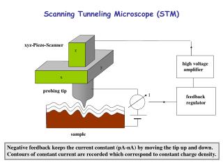
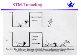
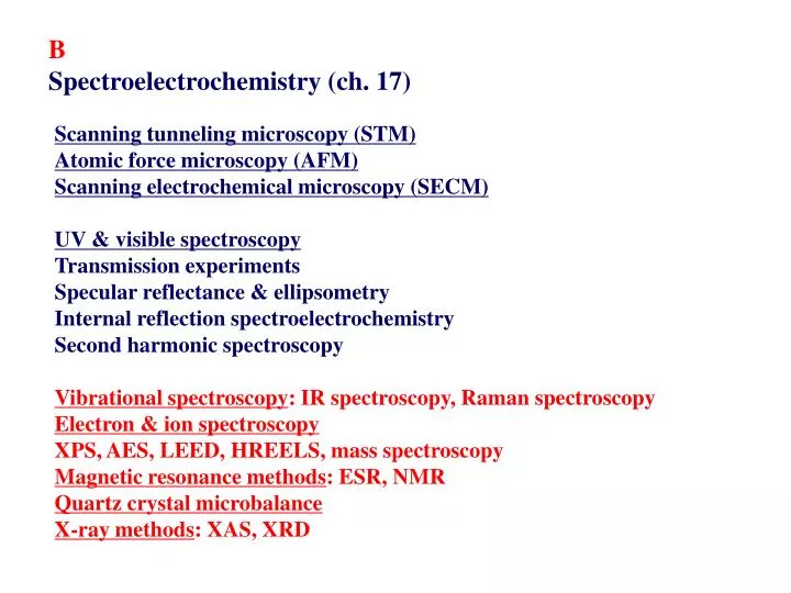
B Spectroelectrochemistry (ch. 17). Scanning tunneling microscopy (STM) Atomic force microscopy (AFM) Scanning electrochemical microscopy (SECM) UV & visible spectroscopy Transmission experiments Specular reflectance & ellipsometry Internal reflection spectroelectrochemistry

E N D
B Spectroelectrochemistry (ch. 17) Scanning tunneling microscopy (STM) Atomic force microscopy (AFM) Scanning electrochemical microscopy (SECM) UV & visible spectroscopy Transmission experiments Specular reflectance & ellipsometry Internal reflection spectroelectrochemistry Second harmonic spectroscopy Vibrational spectroscopy: IR spectroscopy, Raman spectroscopy Electron & ion spectroscopy XPS, AES, LEED, HREELS, mass spectroscopy Magnetic resonance methods: ESR, NMR Quartz crystal microbalance X-ray methods: XAS, XRD
Vibration spectroscopy Infrared spectroscopy Infrared spectroelectrochemistry (IR-SEC)
EMIRS (electrochemically modulated infrared reflectance spectrosocpy) Potential is modulated between one where the species of interest is absent & one where it is electrochemically generated
SNIFTIRS (subtractively normalized interfacial Fourier transform IRS) or PDIRS (potential difference IRS) or SPAIRS (single potential alteration) Spectra obtained separately at two potentials → subtraction
IRRAS (IR reflection absorption spectroscopy) IR absorption at fixed potential
SEIRA (surface enhanced IR absorption) IR to study adsorbed species (reactants, intermediates, products) → orientation & potential dependence of the adsorbed species SNIFTIRS
IR incidence sample ZnSe or KRS-5 crystal In-situ FT-IR spectroscopy • Diffuse Reflectance Infrared Fourier Transform Spectroscopy (DRIFT) • Attenuated Total Reflectance Spectroscopy (ATR) *Transmission measurement *Reflection-Aborption infrared Spectroscopy (RAS) *Photoacoustic Spectroscopy (PAS) *Surface Electromagnetic Wave spectroscopy (SEW)
In-situ FT-IR In situ FT- IR cell
In situFT-IR CO/Pt
Raman spectroscopy: molecular vibrational information complementing IR spec. Raman in electrochemical system: signal enhancement - Resonance Raman spectroscopy (RRS) - Surface enhanced Raman spec. (SERS): molecules adsorbed on certain surfaces (Ag or Au)
Fresh anode In-situ Raman microscopy (탄소재 음극과 LiCoO2양극재 분석) 25oC 60oC LiCoO2 graphite acetylene black Brodd (2003)
Electron and ion spectrometry Ultra high vacuum (UHV) Excitation Detection X-ray photoelectron spectroscopy (XPS) Photons(X-ray) Electrons UV photoelectron spectroscopy (UPS) Photons (UV) Electrons Auger electron spectroscopy (AES) Electrons Electrons Low-energy electron diffraction (LEED) Electrons Electrons High resolution e- E loss spec. (HREELS) Electrons Electrons Rutherford backscattering (RBS) H+ or He+ H+ or He+ Secondary ion mass spec. (SIMS) Ions Ions Laser desorption mass spec. (LDMS) Photons Ions
Detection limits, sampling depth, spot size (spatial resolution)
XPS for copper electrodeposition (a) Bulk Cu (b) Cu UPD
Electrochemical X-ray Photoelectron Spectroscopy Univ. of Illinois
Electrochemical XPS UHV-XPS Glove Box Ex-situ Analysis without Contamination
High resolution electron energy loss spectroscopy • SCN- on Ag(111) • -0.3 V • +0.14 V
Mass spectrometry Differential electrochemical mass spectrometry (DEMS)
DEMS: fuel cell catalysts for methanol(solid) & formic acid(dotted) oxidation
Magnetic resonance methods Electron spin resonance & NMR
X-ray methods Synchrotron X-ray absorption spectroscopy Absorption edge (energy that is just needed to eject a particular core electron, e.g., 1s e- (K edge), 2p3/2 e- (L3 edge) Fe & Fe oxides K-edge: 7.112 keV Within 10-40 eV: X-ray absorption near-edge structure (XANES) (or near-edge absorption fine structure (NEXAFS)) → oxidation state & ligand envirionment About 50 keV: extended X-ray absorption Fine structure (EXAFS) → distance & arrangement of atoms
In-situ XRD In-situ XRD patterns of LixFeSnO4 during initial lithium intercalation and deintercalation.