Download
1 / 40
400 likes | 600 Views
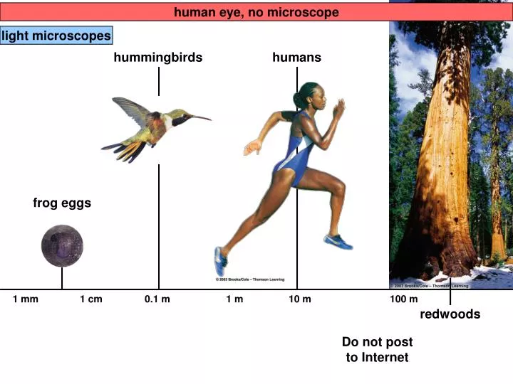
human eye, no microscope. light microscopes. hummingbirds. humans. frog eggs. 1 mm. 1 cm. 0.1 m. 1 m. 10 m. 100 m. redwoods. Do not post to Internet. light microscopes. electron microscopes. lipids. bacteriophages. most animal cells and plant cells. mitochondria, chloroplasts.

E N D
human eye, no microscope light microscopes hummingbirds humans frog eggs 1 mm 1 cm 0.1 m 1 m 10 m 100 m redwoods Do not post to Internet
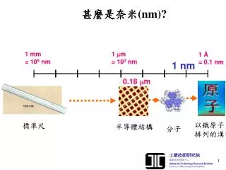
light microscopes electron microscopes lipids bacteriophages most animal cells and plant cells mitochondria, chloroplasts small molecules most bacteria proteins 0.1 nm 1 nm 10 nm 100 nm 1 µm 10 µm 100 µm 1 mm
Impacts, Issues Do not post to Internet
In-text FigurePage 51 Do not post to Internet Rickettsia prowazekii
Diameter (cm): 0.5 1.0 1.5 Surface area (cm2): 0.79 3.14 7.07 Volume (cm3): 0.06 0.52 1.77 Surface- to-volume ratio: 13.17:1 6.04:1 3.99:1
Bacterial cell (prokaryotic) DNA cytoplasm plasma membrane
Animal cell (eukaryotic) DNA in nucleus cytoplasm plasma membrane
Plant cell (eukaryotic) DNA in nucleus cytoplasm plasma membrane
Nucleus: nuclear envelope nucleolus Cytoskeleton: DNA in nucleoplasm microtubules microfilaments intermediate filaments rough ER mitochondrion centrioles smooth ER Golgi body plasma membrane lysosome
lipid bilayer fluid fluid one layer of lipids one layer of lipids
adhesion protein communication protein
recognition protein receptor protein passive transporter active transporters
DNA infolding of plasma membrane
NUCLEUS Chromatin Two membranesof nuclearenvelope Nucleolus Pore ROUGHENDOPLASMICRETICULUM Ribosomes Figure 4.6
Transport vesiclebuds off 4 Ribosome Secretory(glyco-) proteininside transportvesicle Sugarchain 3 Glycoprotein 1 2 ROUGH ER Polypeptide Rough Endoplasmic Reticulum • The rough ER manufactures membranes • Ribosomes on its surface produce proteins Figure 4.8
SMOOTH ER ROUGHER Nuclearenvelope Ribosomes SMOOTH ER ROUGH ER Figure 4.9
The Golgi Apparatus Golgi apparatus Golgiapparatus “Receiving” side ofGolgi apparatus Transportvesiclefrom ER Newvesicleforming “Shipping”side of Golgiapparatus Transport vesiclefrom the Golgi Figure 4.10
Rough ER Transport vesicle(containing inactivehydrolytic enzymes) Plasmamembrane Golgiapparatus Engulfmentof particle Lysosomeengulfingdamagedorganelle “Food” LYSOSOMES Digestion Foodvacuole Figure 4.11B
Figure 3.9a-cPage 46 pore chromatin nucleolus nuclear envelope (two lipid bilayers) cytoplasm ribosome vesicle Do not post to Internet
smooth ER channel, cross-section budding vesicle plasma membrane
Mitochondrion repeated foldings of inner membrane (cristae) outer compartment inner compartment outer membrane inner membrane
MITOCHONDRION Outermembrane Intermembranespace Innermembrane Cristae Matrix Figure 4.16
Figure 3.10(2)Page 48 Do not post to Internet
Plant Cell cell wall chloroplast central vacuole Nucleus: nuclear envelope nucleolus Cytoskeleton: DNA in nucleoplasm microtubules microfilaments rough ER mitochondrion plasmodesma smooth ER Golgi body plasma membrane lysosome-like vesicle
Chloroplast two outer membranes stroma thylakoids
THE CYTOSKELETON AND RELATED STRUCTURES • A network of protein fibers makes up the cytoskeleton Figure 4.17A
Microfilaments, Intermediate Filaments and Microtubules • Microfilaments of actin enable cells to change shape and move • Intermediate filaments reinforce the cell and anchor certain organelles • Microtubules • give the cell rigidity • provide anchors for organelles • act as tracks for organelle movement
Microfilaments, Intermediate Filaments and Microtubules Tubulinsubunit Actin subunit Fibrous subunits 25 nm 7 nm 10 nm MICROFILAMENT INTERMEDIATEFILAMENT MICROTUBULE Figure 4.17B
FLAGELLUM Electron micrograph of sections: Outer microtubule doublet Plasmamembrane Flagellum Centralmicrotubules Outer microtubule doublet Plasmamembrane Basal body Basal body(structurally identical to centriole) Figure 4.18A
Clusters of microtubules drive the whipping action of these organelles Microtubule doublet Slidingforce Dynein arm Figure 4.18B
Cell to Cell Junctions • Tight junctions can bind cells together into leakproof sheets • Anchoring junctions link animal cells • Communicating junctions allow substances to flow from cell to cell
Walls of two adjacent plant cells Vacuole PLASMODESMATA Layers of one plant cell wall Cytoplasm Plasma membrane Figure 4.19A
Tight junctions can bind cells together into leakproof sheets • Anchoring junctions link animal cells • Communicating junctions allow substances to flow from cell to cell TIGHTJUNCTION ANCHORING JUNCTION COMMUNICATING JUNCTION Plasma membranes ofadjacent cells Extracellularmatrix Figure 4.19B