Download
1 / 13
130 likes | 217 Views
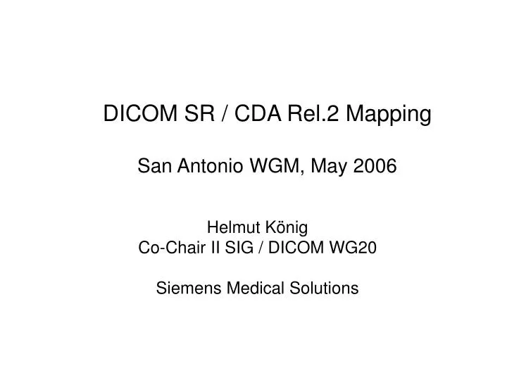
DICOM SR / CDA Rel.2 Mapping San Antonio WGM, May 2006. Helmut König Co-Chair II SIG / DICOM WG20 Siemens Medical Solutions. DICOM SR Report Structure. Header Attributes References: Current Requested Procedure Evidence Sequence Pertinent Other Evidence Sequence Root Content Item

E N D
DICOM SR / CDA Rel.2 MappingSan Antonio WGM, May 2006 Helmut König Co-Chair II SIG / DICOM WG20 Siemens Medical Solutions
DICOM SR Report Structure • Header Attributes • References: • Current Requested Procedure Evidence Sequence • Pertinent Other Evidence Sequence • Root Content Item • Value Type Container • Concept Name Code Sequence -> Report Title • Content Sequence • Content Items (Nodes) • Relationships
Exchange of DICOM SR and CDA Rel.2 Document Contents (1) Mapping Focus: • Start with DICOM SR to CDA Rel.2 Mapping • Inclusion of Imaging Results in Clinical Document as Quoted Excerpts/Extracts + Relevant Context Information • Analyze Delta between Cardiology and Radiology Topics and address both topics
Exchange of DICOM SR and CDA Rel.2 Document Contents (2) Use Cases • Exporting DICOM SR Evidence Document or SR Imaging Report Extracts to CDA Documents • Selected by user and/or automatically for: • Inclusion into clinical document quoting imaging results as part of the clinical evidence • For creation of a CDA summary report on image guided procedures and/or diagnostic imaging results which may be included into the EHR
Echocardiography Use Case (SR Evidence Document) 1) The echocardiography device creates a DICOM SR „Echocardiography Report" containing the findings detected during the examination. The information on the findings comprises coded descriptors, the observation context (e.g. on the human observer), properties of the finding (e.g. numerical measurements) and information on the referenced images. 2) Relevant parts of the „Echocardiography Report" are selected by the verifying human observer (legal authenticator) for inclusion into the clinical document (e.g. for clinical summary report that quotes imaging findings as evidence for conclusions and diagnoses). As a result of this step, the relevant portions of the evidence document are marked/designated. 3) In addition to the marked relevant portions of the evidence document, the relevant document header contents and observation context needed for inclusion into the target document are automatically detected. 4) Selcected portions of the DICOM SR document determined in step 2) and 3) are transformed to the CDA Rel.2 format 5) The transformed document contents are included in the CDA Clinical Document (e.g. clinical summary report) • Transformation from DICOM SR to CDA e.g. by information system capable of receiving DICOM SR documents • Send CDA documents by using HL7 V2 or V3 messages
SR/CDA Mapping Approach • Goal: Determine basic “imaging statements” or “templates” for mapping DICOM SR to CDA Rel.2 • Analysis of SR Content Items and Relationships + Constraints imposed by SR SOP Classes • Analysis of SR Templates such as: • Echocardiography Report • CT/MR Cardiovascular Analysis Report • Vascular Ultrasound Report…
DICOM SR -> CDA Mapping Building Blocks Relevant Document Structures and Contents • Header • Relevant Metadata • Body • Document Structure • Use of Quotations • Observation • Numerical, Nominal, Textual Data • Observation Context • DICOM Composite Object References • Associated Regions of Interest (ROI)
Initial Results & Next Steps • Mapping “Imaging Statements” • Use mapped building blocks for CDA documents and HL7 V3 messages (e.g. Imaging Order, Results Messages) Further Work: • SD TC: Further Discussion on Quoted Contents, Sync on Mapping Approach • DICOM WG10, WG6: Discussion on Reporting Strategy • …
DICOM SR Content Tree (1) Content Items (Nodes) • Value Types: • CONTAINER (Document Structure) • TEXT (Plain Unformatted Text) • CODE (Coded Entries -> Code Sequence Macro) • NUM (Numeric Measurements) • DATE, TIME, DATETIME (Date and Time) • UIDREF (Unique Identifier Reference) • IMAGE (Image Object Reference) • WAVEFORM (Waveform Object Reference) • COMPOSITE (Composite Object Reference) • SCOORD (Spatial Coordinates) • TCOORD (Temporal Coordinates)
DICOM SR Content Tree (2) Content Item Relationships • Relationship Types: • CONTAINS • HAS PROPERTIES • INFERRED FROM • SELECTED FROM • HAS OBS CONTEXT (Has Observation Context) • HAS ACQ CONTEXT (Has Acquisition Context) • HAS CONCEPT MOD (Has Concept Modifier)
HL7 V3 Clinical Document Architecture (CDA) Release 2 • Document Header • Related Acts: Encounter, Order, Parent • Document… • - Participations: PatientRole, Author… • Document Body • Section with Narrative Text • Structured Section Entries Section
Mapping Levels • Header • Section • Document Containers and Codes • Entry • Mapping used for II SIG / WG20 CMETs: