Download
1 / 9
90 likes | 95 Views
The severe acute respiratory syndrome-coronavirus-2-caused coronavirus disease-2019 (COVID-19) has arisen as a serious worldwide public health adversity. Early in the COVID-19 pandemic, an increased incidence of arterial and venous thrombosis was found, linked to systemic inflammation, immobilization, and a prothrombotic environment. Venous thromboembolism (VTE) can manifest itself in a variety of ways. A 55-year-old man presented to the emergency department with peripheral arterial disease (PAD) history.<br><br>Visit Here - https://pubrica.com/services/physician-writing-services/case-report/

E N D
Case report – Sample work A CASE REPORT OF ARTERIAL AND VENOUS THROMBOEMBOLIC ILLNESS IN A COVID-19 PATIENT Copyright © 2022 pubrica. No part of this document may be published without permission of the author
Title: Arterial and venous thromboembolic illness in a COVID-19 patient Summary The severe acute respiratory syndrome-coronavirus-2-caused coronavirus disease-2019 (COVID-19) has arisen as a serious worldwide public health adversity. Early in the COVID-19 pandemic, an increased incidence of arterial and venous thrombosis was found, linked to systemic inflammation, immobilization, and a prothrombotic environment. Venous thromboembolism (VTE) can manifest itself in a variety of ways. A 55-year-old man presented to the emergency department with peripheral arterial disease (PAD) history. The patient had a postoperative cough, dyspnea, chest discomfort, headache, and fever on examination.The patient immediately worsened and was sent to the medium care unit (MCU) for intensive oxygen therapy using a non-rebreathing mask. Despite thromboprophylaxis with low molecular weight heparin, a patient with COVID-19 had an arterial (ischemic stroke) and venous thromboembolic event (PE) after therapy for ischemic stroke with intravenous rt-PA (alteplase) and clopidogrel (LMWH). The patient is still in the MCU but will be transferred to a regular nursing unit soon. Background Due to its quick spread and ease of transmission, the new coronavirus pandemic in the United States has been challenging to comprise. The predominant cause of death with COVID- 19 was pneumonia and respiratory failure, the first focus of investigation and treatment. As the condition advanced and additional instances were discovered, it became apparent that it affects various organs and can have catastrophic implications, including morbidity and mortality. In preliminary trials, pulmonary embolism was an unexpected discovery in COVID-19 patients(Casey et al., 2020). The virus‟s increased systemic inflammatory response, which leads to a greater risk of Hypercoagulability and related thromboembolic illness, is one of the consequences that has since been documented in COVID-19 infection (Oudkerk et al., 2020). Anticoagulants, whether therapeutic or preventive, can be beneficial in preventing and treating the thrombo-inflammatory condition. Providers must, however, first detect the elevated risk of vascular problems and implement severe surveillance procedures. Copyright © 2022 pubrica. No part of this document may be published without permission of the author
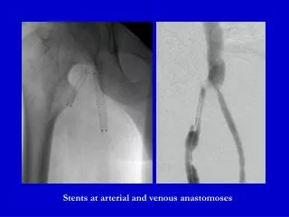
The increased utilization of vascular imaging tests such as CT angiograms and venous duplex ultrasounds was stimulated by the detection of coronavirus-related venous and arterial thromboembolic consequences (Lee et al., 2021). Although thromboprophylaxis with low molecular weight heparin, a patient with COVID-19 had an arterial (ischemic stroke) and venous thromboembolic event (PE) after intravenous rt-PA (alteplase) and clopidogrel treatment for ischemic stroke (LMWH). Furthermore, PE was exposed after two earlier CT pulmonary angiogram (CTPA) imaging examinations had come out negative (Brüggemann et al., 2020). This study examines the incidence of thromboembolic events of COVID-19 patients arterial and venous thromboembolic illness in a COVID-19 patient. Case presentation A 55-year-old male with a history of peripheral arterial disease (PAD) arrived in the emergency department with a 6-day history of increasing cough, dyspnea, thoracic discomfort, headache, and fever. Vital signs revealed temperature, 36.8ºC; heart rate, 123 beats/min; blood pressure, 118/67 mm Hg; respiratory rate, 24 breaths/min; and oxygen saturation, 98% on supplemental oxygen. The lab values for d-dimer (908 g/L), lactate dehydrogenase (585 U/L), and C-reactive protein (53.6 mg/L) were notable. Because of thoracic pain, tachycardia, and an elevated d-dimer level, CTPA was utilized instead of a non-contrast chest CT, which indicated pulmonary abnormalities compatible with COVID-19 pneumonia, whereas pulmonary embolism (PE) was excluded. Upon admission, patients have been given oxygen through a nasal cannula (3 L), amoxicillin, chloroquine, and prophylactic LMWH (nadroparin 3800 IU sc daily). Fig.1 CT scans of the chest (A) At the time of admission, a high-resolution chest CT scan revealed ground-glass anomalies with and without reticulation (“crazy paving”) in a largely peripheral distribution, consistent with COVID-19-related pneumonia. (B) A CTPA was conducted at admission and revealed no evidence of pulmonary emboli. Copyright © 2022 pubrica. No part of this document may be published without permission of the author
(C) On day 2, a high-resolution chest CT scan revealed a significant rise in COVID-19- related pulmonary involvement as well as additional regions of consolidation. (D) On the second day, a CTPA was done, which revealed no symptoms of pulmonary embolism. (E) On day 7, a high-resolution chest CT scan revealed that COVID-19-related lung alterations had improved, with fewer widespread abnormalities and indications consistent with organized pneumonia. (F) On day 7, CTPA revealed lobar (arrow) and subsegmental (not shown) pulmonary emboli. A reverse transcriptase-polymerase chain reaction assay of SARS-CoV2-mRNA on day 2 validated COVID-19 with a Ct value of 23. The patient quickly worsened and was sent to the MCU for non-rebreathing oxygen therapy (15 L, FiO2 80 % ). Copyright © 2022 pubrica. No part of this document may be published without permission of the author
After a second CTPA indicated substantial development of ground-glass opacities and chaotic pavement patterns, PE has been ruled out once more. On day 5, the patient started to experience dysarthria, weakness on the left side, and neglect. Although CT imaging of the brain (including CT angiography and CT perfusion) revealed no brain haemorrhage or arterial obstruction, there was a defect right frontal with a significant mismatch between perfusion and vascular volume measures, indicating a right frontal lobe infarction. In the MCU, there was no evidence of atrial fibrillation (AF). He was given intravenous rt-PA and clopidogrel for secondary stroke prevention after 24 hours. Thromboprophylaxis was also raised to an intermediate dosage (nadroparin 5700 IU sc daily) rationally. On day 7, the respiratory condition worsened much further. Due to the recently provided rt-PA, intermediate-dose thromboprophylaxis, clopidogrel, and two recent negative CTPAs, PE was ruled out. Despite this, when a chest x-ray ruled out pleural effusion and pneumothorax and repeated d-dimers were >10,000 μg/L, a third CTPA was conducted. Despite the fact that the COVID19-related lung injury appeared to be recovering, numerous PE in the right pulmonary artery and bilateral (sub)segmental PE were discovered, Tinzaparin 18,000 IU sc daily) was started as a therapeutic LMWH. The patient is still in the MCU healing but will be transferred to a regular nursing unit shortly. Antiphospholipid antibodies (lupus anticoagulant, IgG and IgM for anticardiolipin, and IgG for anti-2-glycoprotein I) were negative, as was a family history of (VTE). Workup for arterial thromboses A hypercoagulable workup was started to look for arterial and venous thromboses. The workup for arterial thromboses included a negative ELISA for heparin-induced thrombocytopenia (optical density = 0.217, normal 0.400), undetectable anticardiolipin antibodies, and average flow cytometry for paroxysmal nocturnal hemoglobinuria. The lupus anticoagulant test came back positive after 10 days but later negative. As a result, the patient did not fulfil the antiphospholipid antibody syndrome criteria. Copyright © 2022 pubrica. No part of this document may be published without permission of the author
The venous thrombosis workup included negative genetic testing for the prothrombin gene mutation and factor V Leiden and average levels of antithrombin III, protein C, protein S, and homocysteine. Disseminated intravascular coagulation was ruled out by an average prothrombin time (PT), partial thromboplastin time (PTT), and fibrinogen level. Discussion COVID-19 appears to be linked to a high risk of thrombosis due to thrombo- inflammation, which is likely caused by several mechanisms still being investigated (Cui et al., 2020). This instance clarifies why the COVID-19 pandemic causes such terrible outcomes in some people (Zhou et al., 2020). First, it reveals an unusually high burden of consecutive thromboembolic events in both the arterial and venous vascular beds, despite thromboprophylaxis and several other anticoagulant treatments, and in the absence of additional risk factors such as atrial fibrillation, family history of VTE, or antiphospholipid antibodies. The presence of a patent foramen ovale was not ruled out as a stroke risk factor. Even though transthoracic echocardiography revealed a structurally normal heart, there was no „bubble- contrast‟ examination. However, according to juvenile stroke guidelines (Zhou et al., 2020), there was no apparent justification to make a contrast examination based on the patient‟s age, calcifications at the level of the carotid bifurcation, or a history of PAD. Second, it emphasizes the diagnostic issues faced by COVID-19 patients, particularly those who decline rapidly. The inability to distinguish between respiratory failure caused by the evolution of lung-tissue abnormalities such as ground-glass opacities or ARDS on the one hand, and PE on the other, has never been seen before. Whereas in most cases, an alternative diagnosis is comforting and decreases the risk of concomitant PE, this is questionable in individuals with COVID-19. Our case shows that COVID-related anomalies and PE can arise simultaneously or within a short period. To execute CTPA a third time, a repeating d-dimer (>10.000 g/L) was finally required. According to a publication from the Netherlands‟ National Institute of Public Health (RIVM), the combination of increased d-dimer levels with clinical deterioration indicates PE (Oudkerk et al., 2020). Elevated D-dimer levels have also been linked to a bad prognosis (Tang et al., 2020). As a result, we believe CTPA should be evaluated at a low threshold in Copyright © 2022 pubrica. No part of this document may be published without permission of the author
patients with unexplained respiratory failure, significantly if d-dimer levels have been progressively increasing. In retrospect, compression ultrasonography of the legs (CUS) may have been conducted before the second or third CTPA (Stals et al., 2020). If CUS had been positive, there would have been an indication for anticoagulant medication already, and another CTPA may have been avoided. It would have also aided in completely comprehending the sequence of events, although deep vein thrombosis is absent in most patients (van Langevelde et al., 2013). The specific mechanism of COVID-19 infection-induced thromboembolic complications is uncertain. Increased procoagulant factors such as factor VIII, von Willebrand factor, fibrinogen, and a high inflammatory state linked to the cytokine storm leading to fibrinolysis and coagulation activation are probable contributors to COVID-19-related Hypercoagulability (Panigada et al., 2020). In conclusion, Hypercoagulability is a substantial factor in COVID-19-related problems in this case. It indicates that traditional diagnostic and treatment techniques may be insufficient to lower the risk of thromboembolic events, highlighting the need of monitoring COVID-19 patients for these complications. Copyright © 2022 pubrica. No part of this document may be published without permission of the author
References Brüggemann, R., Gietema, H., Jallah, B., ten Cate, H., Stehouwer, C. & Spaetgens, B. (2020). “Arterial and venous thromboembolic disease in a patient with COVID-19: A case report”, Thrombosis Research, 191. 153–155. Casey, K., Iteen, A., Nicolini, R. & Auten, J. (2020). “COVID-19 pneumonia with hemoptysis: acute segmental pulmonary emboli associated with novel coronavirus infection”, The American journal of emergency medicine, 38 (7). 1544-e1. Cui, S., Chen, S., Li, X., Liu, S. & Wang, F. (2020). “Prevalence of venous thromboembolism in patients with severe novel coronavirus pneumonia”, Journal of Thrombosis and Haemostasis, 18 (6). 1421–1424. van Langevelde, K., Sramek, A., Vincken, P.W.J., van Rooden, J.-K., Rosendaal, F.R. & Cannegieter, S.C. (2013). “Finding the origin of pulmonary emboli with a total-body magnetic resonance direct thrombus imaging technique”, Haematologica, 98 (2). 309–315. Lee, E., Krajewski, A., Clarke, C., O‟Sullivan, D., Herbst, T. & Lee, S. (2021). “Arterial and venous thromboembolic complications of COVID-19 detected by CT angiogram and venous duplex ultrasound”, Emergency Radiology, 28 (3). 469–476. Oudkerk, M., Büller, H.R., Kuijpers, D., van Es, N., Oudkerk, S.F., McLoud, T., Gommers, D., van Dissel, J., ten Cate, H. & van Beek, E.J.R. (2020). “Diagnosis, Prevention, and Treatment of Thromboembolic Complications in COVID-19: Report of the National Institute for Public Health of the Netherlands”, Radiology, 297 (1). E216–E222. Panigada, M., Bottino, N., Tagliabue, P., Grasselli, G., Novembrino, C., Chantarangkul, V., Pesenti, A., Peyvandi, F. & Tripodi, A. (2020). “Hypercoagulability of COVID‐19 patients in intensive care unit: A report of thromboelastography findings and other parameters of hemostasis”, Journal of Thrombosis and Haemostasis, 18 (7). 1738–1742. Copyright © 2022 pubrica. No part of this document may be published without permission of the author
Stals, M.A.M., Klok, F.A. & Huisman, M. V. (2020). “Diagnostic management of acute pulmonary embolism in special populations”, Expert Review of Respiratory Medicine, 14 (7). 729–736. Tang, N., Li, D., Wang, X. & Sun, Z. (2020). “Abnormal coagulation parameters are associated with poor prognosis in patients with novel coronavirus pneumonia”, Journal of Thrombosis and Haemostasis, 18 (4). 844–847. Zhou, F., Yu, T., Du, R., Fan, G., Liu, Y., Liu, Z., Xiang, J., Wang, Y., Song, B., Gu, X., Guan, L., Wei, Y., Li, H., Wu, X., Xu, J., Tu, S., Zhang, Y., Chen, H. & Cao, B. (2020). “Clinical course and risk factors for mortality of adult inpatients with COVID-19 in Wuhan, China: a retrospective cohort study”, The Lancet, 395 (10229). 1054–1062. Copyright © 2022 pubrica. No part of this document may be published without permission of the author