Download
1 / 53
530 likes | 540 Views
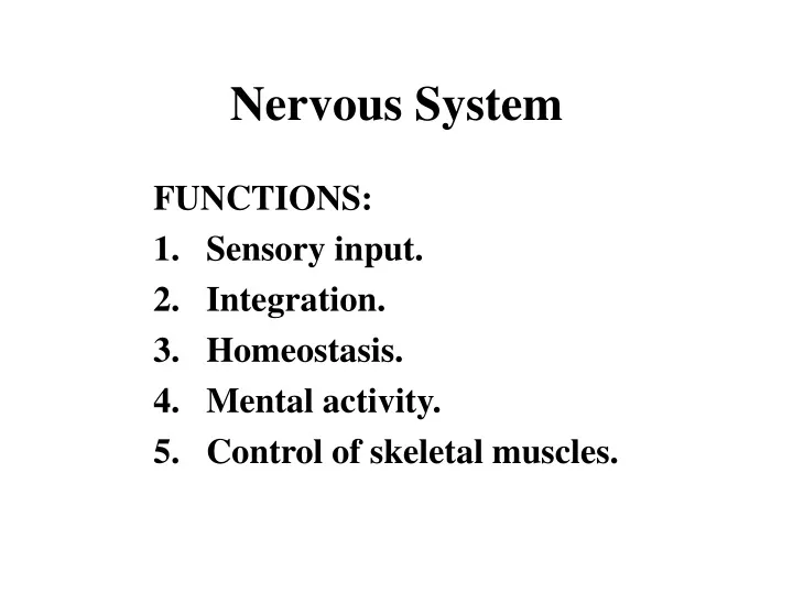
Nervous System. FUNCTIONS: Sensory input. Integration. Homeostasis. Mental activity. Control of skeletal muscles. The Nervous System. Organization of the Nervous System. Central nervous system (CNS) Brain and spinal cord Peripheral nervous system (PNS) Neurons outside the CNS

E N D
Nervous System FUNCTIONS: • Sensory input. • Integration. • Homeostasis. • Mental activity. • Control of skeletal muscles.
Organization of the Nervous System • Central nervous system (CNS) • Brain and spinal cord • Peripheral nervous system (PNS) • Neurons outside the CNS • Sensory division • Afferent fibers transmit impulses from receptors to CNS • Motor division • Efferent fibers transmit impulses from CNS to effector organs
Relationship between motor and sensory fibers of the PNS and the CNS
Autonomic Nervous System • Sympathetic • Fight or flight, stress • Excitatory effects elicited by norepinephrine activating beta receptors • Inhibitory effects elicited by activation of alpha receptors
Parasympathetic • Rest and digest • Digestive system activated, heart rate inhibited, blood vessels dilated • Vagus nerve primarily responsible for activating parasympathetic responses
Synapse Specialized site of intercellular communication. 3 Components: 1. Presynaptic terminal 2. Synaptic cleft 3. Postsynaptic membrane
Neuroglia • Accessory cells of the nervous system • Astrocytes • Support tissue in the CNS form blood-brain barrier • Ependymal • Produce and move cerebral spinal fluid • Microglia • Remove cell debris and bacteria from CNS • Oligodendricytes and Schwann cells • Provide insulation around axons of CNS and PNS neurons
Membrane Potentials • Nervous system functions by establishing concentration gradients and electrical potentials across the membranes • The resting membrane potential of a neuron is negative and is said to be polarized • These gradients are maintained by the sodium potassium pump
Action Potentials • Muscle and nerve cells are exciteable • When a muscle or nerve cell is stimulated Na+ channels open and Na+ rushes into the cell • This causes a local potential • This local potential may not result in action potential • Doesn’t cross the threshold
If the stimulus is sufficient to cause the local potential to cross the threshold an action potential results • The action potential is the complete depolarization of the cell • The action potential is an all-or-nothing event • If the local potential meets threshold, the cell totally depolarizes and the action potential results • If the potential does not meet threshold, no action potential results
Action Potential Propogation • Unmyelinated neurons propogate signals more slowly than myelinated neurons • Myelination acts as an insulator • Electrical signal will jump from node of Ranvier to node of Ranvier • This is called saltatory conduction • Requires less energy than direct propogation
Synapse • Electrical --rare • Chemical --communication occurs in one direction: presynaptic membrane to postsynaptic membrane --action potential is not always propagated.
Synapse Synapses may occur: • neuron to neuron • neuron to another type of cell (neuroeffector) --neuromuscular junction --neuroglandular junction
The Synapse Fig. 8.13
Neurotransmitters --packaged in synaptic vesicles. Nerve endings of the ANS secrete: • Acetylcholine (ACh)--Cholinergic neuron • Parasympathetic effector • Norepinephrine (NE)--Adrenergic neuron • Sympathetic effector
Neurotransmitters diffuse across the synaptic cleft and bind to receptor on the post-synaptic membrane • This can cause membrane channels (Na+, K+, or Cl-) to open or close depending on the neurotransmitter • If stimulatory, Na+ channels will open • If inhibitory, K+ or Cl- channels will open • Cell becomes more negative, hyperpolarized
Receptors 2 types of cholinergic receptors: • Nicotinic • Preganglionic sympathetic and parasympathetic • Muscarinic • parasympathetic 2 types of adrenergic receptors: • Alpha • Generally inhibitory • Beta • Generally excitatory
Autonomic Reflex Arc 1. Receptor 2. Sensory neuron 3. Association neuron 4. Autonomic motor neuron 5. Visceral effector
Adult: Brainstem --medulla oblongata --pons --midbrain Diencephalon --thalamus --hypothalamus --epithalamus Cerebrum Cerebellum Central Nervous System
Brainstem • Medulla oblongata • Inferior portion • Regulation of heart rate, venoconstriction, ventilation, swallowing, , etc.. • Pons • Superior to medulla • Bridge between cerebrum and cerebellum • Midbrain • Audio and visual processing
Cerebellum • Integrates motor signals from cerebral cortex with feedback from PNS • Proprioception • Learning tasks
Dienchephalon • Thalamus • Sensory input from PNS passes through thalamus (relay station) • Epithalamus • Pineal gland – sleep cycle, puberty • Hypothalamus • Master gland • Attached to pituitary by infundibulum • Controls much of homeostasis by stimulating or inhibiting pituitary
Brain Protection: • cranial bones • cranial meninges • cerebrospinal fluid • neuroglia (astrocytes)
CEREBRUM • Largest part of the brain; thinking part • Markings: Gyrus (gyri)-- wrinkle, raised area Fissure(s)-- deep, wide groove(s) Sulcus (sulci)-- shallow groove(s)
CEREBRUM Lobes: 1) Frontal 2) Parietal 3) Temporal 4) Insular 5) Occipital
CEREBRUM Displays lateralization: • left hemisphere language; math/science; reason • right hemisphere music/art; spatial relations; insight/imagination
CEREBRUM • sensory areas • motor areas • association areas
white matter (myelin) dorsal column ventral column lateral column gray matter (non-myelin) posterior horn ventral horn lateral horn Spinal Cord-- Composition
Spinal Cord-- White Matter • myelinated axons that travel along the spinal cord. Ascending--up cord to higher levels Descending-- down cord from brain Across the cord
Spinal Cord • Dorsal roots (sensory) • Ventral roots (motor) combine to form spinal nerve. • Dorsal Root Ganglion