Download
1 / 37
1.29k likes | 4.93k Views
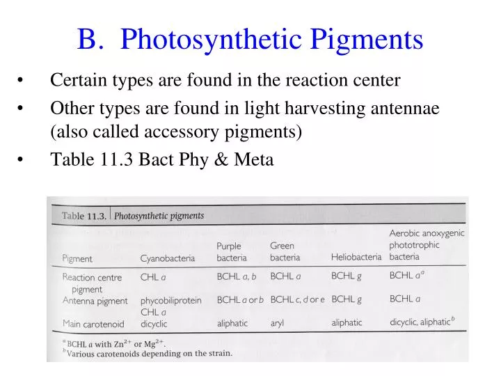
B. Photosynthetic Pigments. Certain types are found in the reaction center Other types are found in light harvesting antennae (also called accessory pigments) Table 11.3 Bact Phy & Meta. B. Photosynthetic Pigments. 1. Chlorophyll and bacteriochlorophyll Pigment found in Reaction center

E N D
B. Photosynthetic Pigments • Certain types are found in the reaction center • Other types are found in light harvesting antennae (also called accessory pigments) • Table 11.3 BactPhy & Meta
B. Photosynthetic Pigments 1. Chlorophyll and bacteriochlorophyll • Pigment found in Reaction center • Also found in light harvesting antennas • Chlorophyll a found in cyanobacteria and eukaryotic algae and plants • Bacteriochlorophyll • Found in other types of phototrophic bacteria • Several different types • An organism may have more than one type
1. Chlorophyll and bacteriochlorophyll Structure • Similar to cytochrome with a tetrapyrrole ring except with a magnesium (or zinc) ion in center • Zinc seen in Bchl a of aerobic anoxygenic bacteria
1. Chlorophyll and bacteriochlorophyll • Side chains off ring vary in different types Fig 17-4 Brock See also Table 11.4 BactPhy & Meta
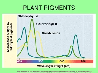
1. Chlorophyll and bacteriochlorophyll • Chlorophyll absorbs in 300-400 and 700 nm • bacteriochlorophylls can absorb in 800 nm rang (near infra red) Fig 17.3 Brock Green line – chlorophyll a of green alga Red line – bacteriochloro-phyll a of purple non-sulfur bacteria
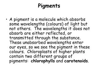
B. Photosynthetic Pigments 2. Carotenoids • Light harvesting accessory pigment – transfers light energy to reaction center (chlorophyll) • Found in all photosynthetic organisms • Absorb in 400-600 nm range • Long isoprenoid structure • Also protect organisms against photooxidation (quench toxic oxygen species) Fig 17.8 Brock Structure of a typical carotenoid
B. Photosynthetic Pigments Structure of a typical phycobilin 3. Phycobilins or phycobiliproteins– • Four linear pyrrole rings bound to a protein • Found in cyanobacteria and red algal chloroplasts • Main light harvesting pigment of these org. • Absorbs in 500-600 nm range
B. Photosynthetic Pigments 4. Pheophytin – • Tetrapyrrole rings of chlorophyll or bacteriochlorophyll without a metal ion in it • Serve as electron carrier in reaction centers • Found in bacteria with pheophytin-quinone type reaction center (cyanobacteria, purple, FAPB and AAPB)
B. Photosynthetic Pigments • Bacteria differ in their absorption spectra based on their pigments • Plants absorb blue and red light and reflect green light Fig 11.4 BactPhy & Meta
C. Phototrophic Bacteria Table 5.1 White
C. Phototrophic Bacteria Table 11.2 BactPhy & Meta
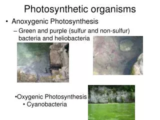
C. Phototrophic Bacteria 1. Cyanobacteria • Found in fresh and marine water and terrestrial habitats, common, abundant • Was called blue-green algae • Photosynthesis similar to algae and plants (chloroplast from an ancestral cyanobacteria) • Electron donor is H2O and make O2 • Carbon from CO2 via Calvin cycle • Pigments in specialized thykaloid membranes • Two photosystems (electrons transport chains)
C. Phototrophic Bacteria 2. Purple bacteria • Purple Sulfur • Found in areas of lakes and oceans and hot springs that are anaerobic and have high sulfides levels (often due to activity of sulfate-reducing bacteria in sediment) • Get electrons from sulfur (H2S), organics or H2 instead of H2O and oxidize sulfur it to S (most accumulate S in granules inside cell) • May also grow as photoheterotrophs but not as chemotroph (aerobic dark growth)
C. Phototrophic Bacteria 2. Purple bacteria B. Purple Non-Sulfur • Found in lake and ponds with low sulfide content • Diverse group spread across proteobacteria • Versatile metabolism (photoautotrophs or photoheterotrophs in anaerobic conditions, chemoorganotrophs in aerobic conditions) • Use H2 or organics as electron donor instead of water for photoautotrophy
C. Phototrophic Bacteria 3. Green bacteria A. Sulfur – • Strict anaerobes • Electron donor is H2S, S, or H2 • Found in anaerobic (anoxic) aquatic environments where H2S is abundant and with low light levels (deep depths) • Fix CO2 by reductive TCA • Have iron-sulfur reaction center with no pheophytin
C. Phototrophic Bacteria 4. Heliobacteria • Only photoheterotrophs - Do not fix CO2 • Obligate anaerobes • Have unique bacteriochlorophyll (g) • No specialized membranes for photosynthesis – pigments in cell membrane
C. Phototrophic Bacteria 5. Aerobic anoxygenic phototrophic bacteria (AAPB) • ~10% of bacteria in upper open ocean • Lack specialized membranes which house photosynthetic apparatuses • Antennae and reaction centers in cell membrane • Bacteriochlorophylla in reaction center • Quasi photosynthetic organisms
D. Photosynthetic Apparatus • How are the pigments organized in cells to capture light energy?
D. Photosynthetic Apparatus Cyanobacteria 1. Cyanobacteria – • Pigments in intracellular thylakoid membranes • May form sacs Chlor-oplast
D. Photosynthetic Apparatus 1. Cyanobacteria – • Thylakoid have phycobilisomes (light capturing accessory structure) • Stick out from membrane • Associated with a reaction center Fig 11.5 BactPhy & Meta
D. Photosynthetic Apparatus 2. Purple bacteria • Have invaginations of cell membrane which can form stacked structures (Fig 11.7 BactPhy and Meta) Fig 5.16 White
D. Photosynthetic Apparatus 3. Green bacteria • Have chlorosome membrane structures with light harvesting pigments • Linked with cell membrane Fig 17.7 Brock Fig 5.16 White
D. Photosynthetic Apparatus 3. Green bacteria • Chlorosome structure serves as one big light harvesting apparatus – • Cell membrane has reaction center with BCHL a • Best for low light levels Fig 11.6 BactPhy & Meta
E. Energy Capture • Mechanisms by which energy is captured as ATP and reduced NAD(P)H • Use electron transport chains • Two ways • Cyclic – electrons not lost, usually only generates proton gradient which can be used for ATP synthesis • Non-cyclic – electrons taken from something and given to different electron acceptor such as NAD(P)+ (means to generate NAD(P)H)
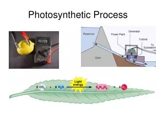
Energy Capture 2. Oxygenic • Electron from water gets excited by light energy and then is passed to electron transport chain • Ultimately can be given to NADP+ 2 H+ out → ATP synthesis
Energy Capture 2. Oxygenic ATP Generation • Can occur to two ways • Non-cyclic phosphorylation • Get electrons from water • Both photosystems involves • Generates both ATP and NADPH • From 2 electrons get 1 ATP and 1 NADPH • Cyclic phosphorylation – dashed arrow • Just photosystem I, • Just generate ATP • Electrons stay in system and not given to NADP+
E. Energy Capture • Anoxygenic cyclic pathway of purple bacteria • Electrons cycle and protons are pumped out LH – light harvesting pigments RC – reaction center Bph – bacteriopheophytin – lacks Mg ion Q – quinone bc1 – cytochrome Fe-S – iron-sulfur protein
E. Energy Capture • Anoxygenic pathway of purple bacteria • To generate NADPH proton motive force is used to run electron transport backwards and give electrons to NADP+ • Several molecules can serve as electron donors 2 H+ out → ATP synthesis P870 – reaction center Bph – bacteriopheophytin – Q – quinone Cyt- cytochrome
Energy Capture 2. Anoxygenic cycle of green sulfur bacteria • cyclic pathway H2S + NAD+ + ADP + P S + NADH + H+ + ATP Fig 11.10 White
F. Carbon Fixation • Incorporation of carbon from CO2 or other 1 carbon molecules into organic molecules • Source of carbon can be • CO2 • Autotrophs – Section 10.8 • Methane (CH4), methanol (CH3OH) or methylamine (CH3NH2)– • methyltrophs – • See section 11.5, 10.8 in BactPhy & Meta
F. Carbon Fixation Ways that CO2 is fixed in Autotrophic Bacteria Can be photo- or chemo- autotrophs • Calvin cycle • Used by photosynthetic eukaryotes and most photosynthetic bactera, cyanobacteria and chemoautotrophs • Acetyl-CoA pathway • Methanogenicarchaea, some sulfate-reducing bacteria, acetogens • Reductive carboxylic acid pathway • Green sulfur photosynthetic bacteria and some archae
PGA 1. Calvin Cycle RuBP • CO2 combines with C5ribulosebiphosphate to make C3 PGA • PGA oxidized to PGALD • Sugar rearrangements generates Fructose 6P and C5RuMP which is converted to RuBP RuMP PGALD PGALD
Calvin Cycle Transfer 2 C • Sugar rearrangements (reactions 4-11) similar to pentose phosphate pathway (transfer 2 or 3 carbon units) C4 C3 C6 Reactions 4-11 are sugar rearrangements C3 C7 C5 C7 C3 Transfer 2 C C5 C5 Fig 13.3 White
1. Calvin cycle • Fig 17-21 Unique or key reactions of Calvin cycle • Most of the reactions are reactions of glycolysis or pentose phosphate pathway
1. Calvin cycle • Integration of Calvin cycle with HMP pathway
F. Carbon Fixation 2. Acetyl-CoA pathway • Used by chemolithotrophs • Assimilation of CO2 by methanogens Fig 10.9
F. Carbon Fixation 3. Reductive TCA cycle • Used by green sulfur bacteria and some chemolitho-trophs • Run TCA backwards – need 3 needs enzymes for steps 3, 5, 8 8 Fig 10.9