Download
1 / 57
570 likes | 747 Views
CASE PRESENTATION ON. CEREBRO VASCULAR ACCIDENT. Name: Mr. X Case number:173815 Age:89 Gender: Male Nationality: Saudi Date of Admission:03/06/2013 Complaints :Left side weakness, slurred speech, facial droop on left side, SOB. General Appearance : Bedridden Conscious Weak-looking

E N D
CASE PRESENTATION ON CEREBRO VASCULAR ACCIDENT
Name: Mr. X • Case number:173815 • Age:89 • Gender: Male • Nationality: Saudi • Date of Admission:03/06/2013 • Complaints :Left side weakness, slurred speech, facial droop on left side, SOB
General Appearance: • Bedridden • Conscious • Weak-looking • Facial droop present on lt side. • Pale in appearance • Thin
Vital Signs: • Blood Pressure: 130/80 mmHg • Pulse Rate: 89 bpm • Respiratory Rate: 23 bpm • Temperature: 37.0°c Skin: • Blister noted on buttocks • Warmth to touch • Poor skin turgur • Wrinkly skin • Scaly
Head • Skull is smooth, no depression. • Loss of hair on scalp. Neck • No lymph node enlargement present. • No masses and leison seen. Thorax: • Symmetrical though the ribs are prominent. • Bilateral chest movement present • wheezing and crackles present . • Dysponea present . Abdomen: • Rigid, with active peristalsis.
Genital Area: • Minimal pubic hair. No hernia noted. Muscular skeletal • Nail beds are thick and dry. • Unable to move both extremitties. • With non-pitting edema on both feet, grade1. • Cannot perform ADL.
PAST MEDICAL HISTORY: PATIENT HAS THE HISTORY OF CVA(LEFT),HYPERTENSION,DM-II, SINCE 5YEARS. HE IS ON MEDICATION . • SUCH AS , TAB AMLOR 5 MG BD TAB GLYSIPHAGE 500 MG BD
PRESENT MEDICAL HISTORY THE PATIENT X IS BROUGHT INTO ER BY SAUDI REDCRESENT AMBULANCE WITH THE CHIEF COMPLAINTS OF LEFT SIDE WEAKNESS ,SLURRED SPEECH ,FACIAL DROOP ON LEFT SIDE AND SHORTNESS OF BREATHING. THE PATIENT THOROUGHLY EXAMINED BY ER DOCTOR. CT BRAIN WITHOUT CONTRAST HAS DONE .ECG IS DONE AND ALL LAB INVESTIGATIONS ARE SENT. PATIENT DIAGNOSED AS RIGHT SIDE ISCHEMIC STROKE AND ADMITTED IN ICU FOR FURTHER MANAGEMENT SURGICAL HISTORY:PATIENT HAS NO PRESENT AND PAST SURGICAL HISTORY
CT Scan of brain without contrast: FINDINGS: The brain is morphologically normal. No acute hemorrhage. IMPRESSION: Diffuse cerebral atrophy with small periventricular white matter chronic ischemic changes.
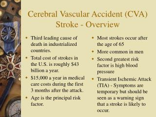
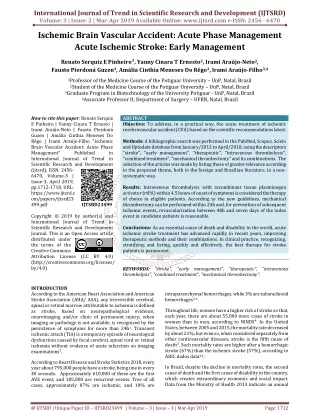
INTRODUCTION: Cerebro vascular accident or brain attack is the onset and persistence of neurologic dysfunction lasting longer than 24 hours and resulting from disruption of blood supply to the brain and indicates infraction rather than ishchemia.stroke are classified as ischemia and hemorrhagic.
DEFINITION; Cerebrovascular accident: The sudden death of some brain cells due to lack of oxygen when the blood flow to the brain is impaired by blockage or rupture of an artery to the brain. A CVA is also referred to as a stroke.
Ischemic stroke. About 85 percent of strokes are ischemic strokes. The most common ischemic strokes include: • Thrombotic stroke. • Embolic stroke. • Hemorrhagic stroke. Hemorrhagic stroke occurs when a blood vessel in your brain leaks or ruptures.. • Transient ischemic attack (TIA). A transient ischemic attack (TIA) — also called a ministroke — is a brief episode of symptoms similar to those you'd have in a stroke. A transient ischemic attack is caused by a temporary decrease in blood supply to part of your brain. TIAs often last less than five minutes.
ANATOMY AND PHYSIOLOGY The brain is one of the largest and most complex organs in our body. It controls our body, receives information, analysis information and stores information. It is made up of more than 100 billion nerves that communicates in trillions of connections called synapse. The skull consisting of 22 bones all together. These bones are divided into 8 cranial bones and 14 facial bones. Cranial bones form the cranial cavity and protects the brain.
ANATOMY AND PHYSIOLOGY CRANIAL NERVES Olfactory : sense of smell. Optic Nerve : sight of retina. Oculomotor Nerve : eye movement and pupil constriction. Trochlear Nerve: eye movements. Trigeminal Nerve : carry somatosensory information to face, head and chewing muscles of jaws. Abducens Nerve : eye movement. Facial : control the muscles used for facial expressions (smiling, frowning etc). It also stimulates salivary glands to produce saliva.
ANATOMY AND PHYSIOLOGY Vestibulocochlear VIII: hearing and balance. Glossopharyngeal IX: taste sensation ,gag reflexes. Vagus X: It carries somatosensory information from organs of thoracic, abdominal cavity including heart and from that of gastrointestinal tract. Spinal Accessory Nerve XI: leads to muscles of neck, back and larynx. It controls the head movement. Hypoglossal Nerve XII:controls the muscles of tongue
ANATOMY AND PHYSIOLOGY Meninges Meninges are the connective tissue membrane enclosing the brain and the spinal cord. It is divided into 3.outer most duramater,arachanoid mater and the inner most piamater.
ANATOMY AND PHYSIOLOGY Lobes of brain Frontal lobe: is responsible for problem solving,judgement and motor function. Parietal lobe: manage sensation, hand writing and body position. Temporal lobe: is involved with memory and hearing. Occipital lobe: contain the brains visual processing system.
ANATOMY AND PHYSIOLOGY Cerebrum: Cerebrum is the most superior part of the brain. It is made up of by thick gray matter as surface layer and internally with white matter.It consist of thalamus, hypothalamus and epithalamus.
ANATOMY AND PHYSIOLOGY Cerebellum: Cerebellum located dorsal to the pons and medulla. It receives the impulses from cerebral motor cortex, various stem and sensory receptors in order to control skeletal muscle contraction.
Brain stem: Brain stem is similarly structured as the spinal cord. It is divided in to midbrain ,pons and medulla oblongata.mid brain acts as a fiber pathway between higher and lower brain centres.The pons mainly a conduction region also contribute to the regulation of respiration and cranial nerves. Medulla oblongata regulate the respiratory rhythm, heart rate,B P etc...
BLOOD SUPPLY TO THE BRAIN The brain receives about 25% of the body's oxygen, but it cannot store it. Brain cells require a constant supply of oxygen to stay healthy and function properly. Therefore, blood needs to be supplied continuously to the brain through two main arterial systems: The carotid arteries come up through either side of the front of the neck. (To feel the pulse of a carotid artery, place your fingertips gently against either side of your neck, right under the jaw.) The basilar artery forms at the base of the skull from the vertebral arteries, which run up along the spine, join, and come up through the rear of the neck.
ETIOLOGY • Partial or complete occlusion of a cerebral blood flow to an area of the brain due to; a. Thrombous. b. Embolus. • Aneurysm: a balloon-like bulge or weakening of an artery wall that ruptures, releasing blood into the subarachnoid space around the brain.. • Head injury. • Atrial fibrillation.
HEAD TRAUMA OR A RUPTURED ANEURYSM THROMBOSIS ,EMBOLISM,OR LACUNAR INFRACT HYPERTENSION PATHO PHYSIOLOGY UN CONTROLLED BLEEDING BETWEEN THE INNERMOST TWO OF THE THREE MENINGES,THE PIAMATTER AND ARACHANOID MATTER. LOSS OF PERFUSION TO AN AREA OF THE BRAIN -ISHCHEMIA ARTERY INSIDE THE BRAIN FAIL TO CONSTRICT BLOOD POOLS IN THE SUBARACHANOID SPACE-SUBARACHANOID HAEMORRHAGE BURST AND BLEED-INTRA CEREBRAL HAEMORRHAGE IN ABILITY TO MOVE ,UNDERSTAND ,FORMULATE SPEECH OR SEE IN ONE SIDE OF THE VISUAL FIELD. CVA
THERAPUTIC TREATMENT • Support of vital function • Maintain airway, breathing oxygenation and circulation • Neurological assessment • To check the deterioration related to re bleed or development of cerebral edema. • Neurological consultation for possible evacuation of intracranial hemorrhage may be explored. • Reversal of coagulopathies: • Anticoagulant therapy if thrombus or embolus is present, antiplatelet therapy. • If ischemic type, thrombolytic therapy with recombinant tissue plasminogen activator, (t-PA) with in 3 hours of onset.
TREATMENT • Management for BP with in prescribed parameter. • Maintain BP less than 160 /90 to reduces vessel wall stressors. • Prophylactic treatment of seizure with phenytoin. • Anti-inflammatory or osmotic diuretics may be used to reduce cerebral edema and intracranial pressure. • Evaluate any signs of increased intracranial pressure. • IV fluid at maintaince until able to tolerate oral feed • SURGICAL MANAGEMENT • Hemicraniectomy • Carotid endraterectomy • Also called stent placement( may be done prophylactically) to improve cerebral flow when carotid arteries are narrowed by arteriosclerotic plaque.
Aspiration pneumonia • Decubitus ulcers • Dementia • Disability: • Difficulty speaking • Difficulty swallowing • Arm weakness (unilateral) • Leg weakness (unilateral) • Facial weakness • Inability to live independently • Urinary incontinence • Bowel incontinence • Memory loss • Muscle spasms • Osteoporosis • Chronic pain • Recurrent stroke • Tremor
Nursing Intervention includes: • Monitor Vital Signs, especially the blood pressure • Monitor the Neurovascular Status • Proper positioning. • Give due medications on time • Institute safety and aspiration precautions • Main tain skin integrity • Helps the patient to do ADL activities • Prevent patient from falls.
ACTIVITY AND REST; • Disturbed sleeping pattern • Impaired physical mobility • Risk for activity intolerance. BREATHING • Ineffective Airway Clearance • Ineffective Breathing Pattern FATIGUE • Ineffective Breathing Pattern • Decreased Cardiac Output • Risk for Decreased Cardiac Tissue Perfusion • Self Care Deficit