Download
1 / 49
530 likes | 679 Views
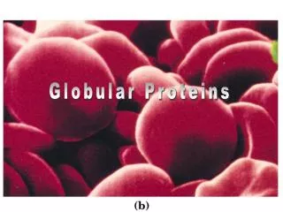
Globular proteins. Globular proteins. Proteins are biochemical compounds consisting of one or more polypeptides typically folded into a globular or fibrous form in a biologically functional way. Globular proteins.

E N D
Globular proteins • Proteins are biochemical compounds consisting of one or more polypeptides typically folded into a globularor fibrousform in a biologically functional way.
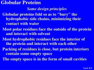
Globular proteins • Globular proteins, or spheroproteins comprising "globe"-like proteins that are more or less soluble in aqueous solutions. • This main characteristic helps distinguishing them from fibrous proteins (the other class), which are practically insoluble.
Globular proteins • Unlikefibrousproteinswhich only play a structural function, globular proteins can act as: • Enzymes. • Messengers, by transmitting messages to regulate biological processes. This function is done by hormones (i.e. insulin etc). • Transportersof other molecules through membranes.
Globular hemeprotein • Hemeproteinsare a group of specialized proteins that contain hemeasa tightly bound prosthetic group. • Prosthetic group is a tightly bound, specific non-polypeptide unit required for the biological function of some proteins. • Theprosthetic group may be organic (such as a vitamin, sugar, or lipid) or inorganic (such as a metal ion), but is notcomposed of amino acids. • The role of the hemegroup is dictated by the environment created by the three-dimensional structure of the protein.
Globular hemeprotein • For example, the hemegroup of a cytochrome functions as an electron carrier that is alternately oxidized and reduced. • In contrast, the hemegroup of the enzyme catalaseis part of the active site of the enzyme that catalyzes the breakdown of hydrogen peroxide . • In hemoglobinand myoglobin, the two most abundant hemeproteins in humans, the hemegroup serves to reversibly bind oxygen.
Structure of heme • Hemeis a complex of protoporphyrin IXandferrousiron (Fe2+). • The iron is held in the center of the hememolecule by bonds to the four nitrogensof the porphyrinring. • The heme(Fe2+) can form two additional bonds, one on each side of the planar porphyrinring.
Myoglobin • For example, in myoglobinand hemoglobin, one of these positions is coordinated to the side chain of a histidine residue of the globin molecule, whereas the other position is available to bind oxygen.
Structure and function of myoglobin • Myoglobinhemeproteinpresent in heart and skeletal muscle, functions both as a reservoir for oxygen, and as an oxygencarrier that increases the rate of transport of oxygenwithin the muscle cell. • Myoglobinconsists of a single polypeptide chain that is structurally similar to the individual subunit polypeptide chains of the hemoglobinmolecule.
1. α-Helical content • Myoglobinis a compact molecule, with approximately 80% of its polypeptide chain folded into eightstretches of α-helix. • These α-helicalregions are terminated either by the presence of proline, whose five-membered ring cannot be accommodated in an α-helix,or by β-bendsand loops stabilized by hydrogenbondsand ionicbonds.
2. Location of polar and nonpolar amino acid residues • The interior of the myoglobinmolecule is composed almost entirely of nonpolaraminoacids. • They are packed closely together, forming a structure stabilized by hydrophobicinteractionsbetween these clustered residues . • In contrast, chargedaminoacids are located almost exclusively on the surface of the molecule, where they can form hydrogenbonds, both with each other and with water.
3. Binding of the heme group • The hemegroup of myoglobinsits in a crevice in the molecule, which is lined with nonpolar amino acids. • Notable exceptions are two histidineresidues . • One, the proximal histidine, binds directly to the iron of heme. • The second, or distal histidine, does not directly interact with the hemegroup, but helps stabilize the binding of oxygento the ferrousiron.
Structure and function of hemoglobin • Hemoglobinis found exclusively in red blood cells, where its main function is to transport oxygenfrom the lungs to the capillaries of the tissues. • Hemoglobin A, the major hemoglobin in adults, is composed of four polypeptide chains ; twoalpha (α) chains and twobeta (β)chains held together by noncovalentinteractions.
Structure and function of hemoglobin • Each subunit has stretches of α-helicalstructure, and a hemebinding pocket similar to that described for myoglobin. • However, the tetramerichemoglobinmolecule is structurally and functionally more complex than myoglobin. • For example, hemoglobincan transport H+ and CO2 from the tissues to the lungs, and carry four molecules of O2 from the lungs to the cells of the body. • Further, the oxygen-binding properties of hemoglobinare regulated by interaction with allosteric effectors .
Quaternary structure of hemoglobin • The hemoglobin tetramer can be envisioned as being composed of two identical dimers (αβ)1, and (αβ)2, in which the numbers refer to dimers one and two. • The two polypeptide chains within each dimer are held tightly together, primarily by hydrophobicinteractions. • Hydrophobicaminoacidresidues are localized not only in the interior of the molecule, but also in a region on the surface of each subunit. • Interchainhydrophobicinteractions form strong associations between α-subunits andβ-subunits in the dimers. • Ionicand hydrogenbonds also occur between the members of the dimer.
Quaternary structure of hemoglobin • In contrast, the twodimers are able to move with respect to each other, being held together primarily by polar bonds. • The weaker interactions between these mobile dimers result in the two dimers occupying different relative positions in deoxyhemoglobinas compared with oxyhemoglobin: • T form • R form
T form • The deoxyform of hemoglobinis called the "T" or taut(tense) form. • In the Tform, the two α βdimers interact through a network of ionic bonds and hydrogen bonds that constrain the movement of the polypeptide chains. • The Tform is the low oxygen-affinity form of hemoglobin.
R form • The binding of oxygen to hemoglobincauses the rupture of some of the ionic bonds and hydrogenbondsbetween the α βdimers. • This leads to a structure called the "R," or relaxed form, in which the polypeptide chains have more freedom of movement. • The Rform is the high oxygen-affinity form of hemoglobin.
Binding of oxygen to myoglobin and hemoglobin • Myoglobincan bind only one molecule of oxygenbecause it contains only one hemegroup. • In contrast, hemoglobincan bind fouroxygenmolecules—one at each of its four hemegroups. • The degree of saturation (Y) of these oxygen-binding sites on all myoglobinor hemoglobinmolecules can vary between zero(all sites are empty) and 100percent (all sites are full).
Oxygen dissociation curve • A plot of Ymeasured at different partial pressures of oxygen (pO2) is called the oxygen dissociation curve. • The curves for myoglobinand hemoglobinshow important differences . • Myoglobinhas a higher oxygen affinity at all pO2 values than does hemoglobin. • The partial pressure of oxygen needed to achieve half-saturation of the binding sites (P50) is approximately 1 mm Hg for myoglobinand 26 mm Hg for hemoglobin. • [Note: The higher the oxygen affinity (that the more tightly oxygen binds), the lower the P50.
a. Myoglobin: • The oxygen dissociation curve for myoglobin has a hyperbolic shape. This reflects the fact that myoglobin reversibly binds a single molecule of oxygen. • Thus, oxygenated (MbO2) and deoxygenated (Mb) myoglobin exist in a simple equilibrium: • The equilibrium is shifted to the right or to the left as oxygen is added to or removed from the system. • [Myoglobinis designed to bind oxygen released by hemoglobinat the low PO2 found in muscle. Myoglobin, in turn, releases oxygenwithin the muscle cell in response to oxygendemand.]
b. Hemoglobin • The oxygen dissociation curve for hemoglobinis sigmoidal in shape, indicating that the subunits cooperate in binding oxygen. • Cooperative binding of oxygen by the four subunits of hemoglobin means that the binding of an oxygen molecule at one hemegroup increases the oxygen affinity of the remaining hemegroups in the same hemoglobin molecule . • This effect is referred to as heme-heme interaction . • Although it is more difficult for the first oxygen molecule to bind to hemoglobin, the subsequent binding of oxygen occurs with high affinity, as shown by the steep upward curve in the region near 20 to 30 mm Hg.
Allosteric effects • The ability of hemoglobin to reversibly bind oxygen is affected by: • The pO2 (through heme-heme interactions) • The pH of the environment. • The partial pressure of CO2 (pCO2). • The availability of 2,3- bisphosphoglycerate. • These are collectively called allosteric("other site") effectors, because their interaction at one site on the hemoglobinmolecule affects the binding of oxygen to hemegroups at other locations on the molecule. • [Note: The binding of oxygen to myoglobinis not influenced by the allosteric effectors of hemoglobin.]
1. Heme-heme interactions: • The sigmoidal oxygen-dissociation curve reflects specific structural changes that are initiated at one hemegroup and transmitted to other hemegroups in the hemoglobintetramer. • The net effect is that the affinity of hemoglobinfor the last oxygenbound is approximately 300 times greater than its affinity for the first oxygenbound.
1. Heme-heme interactions: a. Loading and unloading oxygen: • The cooperative binding of oxygenallows hemoglobinto deliver more oxygento the tissues in response to relatively small changes in the partial pressure of oxygen. • For example, in the lung, the concentration of oxygenis high and hemoglobinbecomes virtually saturated (or "loaded") with oxygen. • In contrast, in the peripheral tissues, oxyhemoglobinreleases (or "unloads") much of its oxygenfor use in the oxidative metabolism of the tissues .
2. Bohr effect • The release of oxygenfrom hemoglobinis enhanced when the pH is loweredor when the hemoglobinis in the presence of an increased partial pressure of CO2 (pCO2). • Both result in a decreasedoxygenaffinity of hemoglobinand, therefore, a shift to the right in the oxygen dissociation curve and both, then, stabilize the T state. • This change in oxygenbinding is called the Bohr effect. • Conversely, raisingthe pH or loweringthe concentration of CO2 results in a greater affinity for oxygen, and a shift to the left in the oxygen dissociation curve and stabilization of the R state.
2. Bohr effect a. Source of the protons that lower the pH: • The concentration of both CO2and H+ in the capillaries of metabolically active tissues is higher than that observed in alveolar capillaries of the lungs, where CO2 is released into the expired air. • In the tissues, CO2 is converted by carbonicanhydraseto carbonic acid: • which spontaneously loses a proton, becoming bicarbonate(the major blood buffer):
2. Bohr effect a. Source of the protons that lower the pH: • The proton produced by this pair of reactions contributes to the loweringof pH. • This differential pH gradient (lungs having a higherpH , tissues a lowerpH) favors the unloadingof oxygenin the peripheral tissues, and the loadingof oxygenin the lung. • Thus, the oxygenaffinity of the hemoglobinmolecule responds to small shifts in pH between the lungs and oxygen-consuming tissues, making hemoglobina more efficient transporter of oxygen.
2. Bohr effect b. Mechanism of the Bohr effect • The Bohr effect reflects the fact that the deoxyform of hemoglobinhas a greater affinity for protons than does oxyhemoglobin . • This effect is caused by ionizable groups, such as specific histidineside chains that have higher pKas in deoxyhemoglobinthan in oxyhemoglobin. • Therefore, an increase in the concentration of protons (resulting in a decreasein pH) causes these groups to become protonated (charged) and able to form ionic bonds (also called salt bridges). • These bonds preferentially stabilize the deoxyform of hemoglobin, producing a decrease in oxygenaffinity.
2. Bohr effect b. Mechanism of the Bohr effect • The Bohreffect can be represented schematically as: • where an increase in protons (or a lower pO2) shifts the equilibrium to the right(favoring deoxyhemoglobin), whereas an increase in pO2 (or a decrease in protons) shifts the equilibrium to the left.
3. Effect of 2,3- Bisphosphoglycerate on oxygen affinity • 2,3-Bisphosphoglycerate (2,3-BPG) is an important regulator of the binding of oxygento hemoglobin. • It is the most abundant organic phosphate in the red blood cell, where its concentration is approximately that of hemoglobin. • 2,3-BPG is synthesized from an intermediate of the glycolytic pathway.
3. Effect of 2,3- Bisphosphoglycerate on oxygen affinity a. Binding of 2,3-BPG to deoxyhemoglobin • 2,3-BPG decreases the oxygen affinity of hemoglobinby binding to deoxyhemoglobinbut not to oxyhemoglobin. • This preferential binding stabilizes the tautconformation of deoxyhemoglobin.
3. Effect of 2,3- Bisphosphoglycerate on oxygen affinity b. Binding site of 2,3-BPG: • One molecule of 2,3-BPG binds to a pocket, formed by the two β-globin chains, in the center of the deoxyhemoglobin tetramer. • This pocket contains several positivelycharged amino acids that form ionic bonds with the negativelycharged phosphate groups of 2,3-BPG. • [Note: A mutation of one of these residues can result in hemoglobin variants with abnormally high oxygen affinity.] • 2,3-BPG is expelled on oxygenation of the hemoglobin.
3. Effect of 2,3- Bisphosphoglycerate on oxygen affinity c. Shift of the oxygen-dissociation curve • Hemoglobinfrom which 2,3-BPG has been removed has a high affinity for oxygen. • However, as seen in the red blood cell, the presence of 2,3- BPG significantly reduces the affinity of hemoglobinfor oxygen, shifting the oxygen-dissociation curve to the right. • This reduced affinity enables hemoglobinto release oxygenefficiently at the partial pressures found in the tissues.
4. Binding of CO2 • CO2 produced in metabolism is hydrated and transported as bicarbonate ion. • However, some CO2 is carried as carbamate bound to the uncharged α-amino groups of hemoglobin (carbamino-hemo-globin which can be represented schematically as follows: • The binding of CO2 stabilizes the T (taut) or deoxyform of hemoglobin, resulting in a decrease in its affinity for oxygen, In the lungs CO2, disassociates from the hemoglobin, and is released in the breath.
5. Binding of CO • Carbon monoxide (CO) binds tightly (but reversibly) to the hemoglobiniron, forming carbon monoxyhemoglobin (HbCO) or carboxyhemoglobin. • When carbon monoxide(CO) binds to one or more of the four hemesites, hemoglobinshifts to the relaxed (R) conformation, causing the remaining hemesites to bind oxygen with high affinity. • This changes the normal sigmoidal shape toward a hyperbola. • As a result, the affected hemoglobin is unable to release oxygen to the tissues
5. Binding of CO • [Note: The affinity of hemoglobin for COis 220 times greaterthan for oxygen. • Consequently, even minute concentrations of COin the environment can produce toxic concentrations of carbon monoxyhemoglobinin the blood. • For example increased levels of COare found in the blood of tobacco smokers. • COtoxicity appears to result from a combination of tissue hypoxia and direct carbon monoxide-mediated damage at the cellular level. • COpoisoning is treated with 100% oxygenat high pressure which facilitates the dissociation of COfrom the hemoglobin
Summary • Hemoglobin A, the major hemoglobin in adults, is composed of four polypeptide chains (two αchains and two βchains, α2β2) held together by noncovalent interactions. • The subunits occupy different relative positions in deoxyhemoglobincompared with oxyhemoglobin. • The deoxyform of hemoglobin is called the “T,” or taut(tense) form. • Ithas a constrained structure that limits the movement of the polypeptide chains. • The Tform is the low-oxygen-affinity form of hemoglobin.
Summary • The binding of oxygento hemoglobincauses rupture of some of the ionicand hydrogenbonds. • This leads to a structure called the “R,” or relaxedform, in which the polypeptide chains have more freedom of movement. • The Rform is the high-oxygen-affinity form of hemoglobin. • The oxygendissociation curve for hemoglobinis sigmoidal in shape (in contrast to that of myoglobin, which is hyperbolic), indicating that the subunits cooperate in binding oxygen. • Cooperative binding of oxygenby the four subunits of hemoglobinmeans that the binding of an oxygenmolecule at one hemegroup increases the oxygenaffinity of the remaining hemegroups in the same hemoglobinmolecule.
Summary • Hemoglobin’sability to bindoxygenreversibly is affected by the pO2(through heme-hemeinteractions), the pH of the environment, the pCO2, and the availability of 2,3-bisphosphoglycerate (2,3-BPG). • For example, the release of O2from Hbis enhanced when the pH is lowered or the pCO2 is increased (the Bohreffect), such as in exercising muscle, and the oxygendissociation curve of Hbis shifted to the right. • To cope long-term with the effects of chronic hypoxia or anemia, the concentration of 2,3-BPGin RBCs increases. • 2,3-BPG binds to the Hband decreases its oxygenaffinity, and it, therefore, also shifts the oxygen-dissociation curve to the right. • Carbon monoxide(CO)binds tightly (but reversibly) to the hemoglobiniron, forming carbon monoxyhemoglobin (HbCO)