Download
1 / 48
480 likes | 647 Views
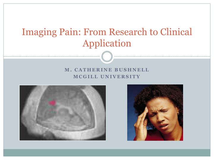
Imaging Pain: From Research to Clinical Application. M. Catherine Bushnell McGill University. Brain imaging allows us to measure neural basis of pain perception. Important technique for examining neural changes related to chronic pain. Brain imaging techniques used to study pain mechanisms

E N D
Imaging Pain: From Research to Clinical Application M. Catherine Bushnell McGill University
Brain imaging allows us to measure neural basis of pain perception Important technique for examining neural changes related to chronic pain
Brain imaging techniques used to study pain mechanisms MRI: provides functional and anatomical information PET: provides neurochemical information Evoked Potentials: provides temporal information
S1 S2 ACC IC Imaging reveals sensory and limbic regions activated by pain ACC: Anterior cingulate cortex; IC: Insular cortex.Apkarian A, et al. Eur J Pain. 2005;9:463–485.
Sensory and limbic regions have different roles in pain processing Pain affect without “pain sensation” in patient with postcentral lesion Ploner et al. 1999
Imaging shows that the pain network activated by many types of pain
Chronic pain can be associated with changes in pain processing. Frida Kahlo
In chronic pain patients the pain network can be activated by tactile stimuli (allodynia) Post-herpetic neuralgia Diabetic neuropathy Back pain
Tactile allodynia related to neuropathic pain reflected in the brain Hofbauer RK, et al. Clin J Pain. 2006;22:104–108.
Chronic pain can alter brain resting state activity Control DMN Patient DMN DMN Patient-Control DMN = Default mode network. Cauda F. PLoS ONE. 2009;4:e4542. diabetic neuropathic pain
Hypersensitivity in “functional” pain syndromes Vulvarvestibulitis Pukall et al 2005
Increased stimulus-evoked brain activation to light touch in vulvar vestibulitis Pukall CF, et al. Pain. 2005;115:118–127.
Hypersensitivity in fibromyalgia Wood et al, Eur. J. Pain 2007
Increased activation to pressure in fibromyalgia Gracely et al 2002
Measuring ongoing chronic pain in MRI scanner Pain intensity = 10/10 Pain intensity = 0/10 MRI: Magnetic resonance imaging. Baliki MN, et al. J Neurosci. 2006;21:12165–12173.
Chronic back pain has transient and sustained components Baliki MN, et al. J Neurosci. 2006;21:12165–12173.
Chronic back pain activates two brain circuits Correlates of increasing pain are similar to acute pain processing Correlates of high sustained pain involve emotional and cognitive regions Baliki MN, et al. J Neurosci. 2006;21:12165–12173.
Imaging shows that some cortical regions are involved in descending pain modulation
Descending modulation of pain Information from cortex ultimately received in spinal cord Schweinhardt and Bushnell, J. Clin. Investigation, in press
Psychological factors modulate pain via these descending modulatory pathways Emotions Attention
Attention to pain Distraction from pain Attention Modulates Pain Bushnell et al. 1999
Good mood + Pain Bad mood + Pain Mood alters pain-evoked activity in limbic brain regions Emotions alters pain Anterior cingulate cortex Villemure & Bushnell 2009
Attention and emotion activate different modulatory circuitry in brain Villemure & Schweinhart 2010
Attentional focussing and/or negative emotional states can contribute to chronic pain states
Major depressive disorder associated with altered descending inhibition during pain Strigo I et al, Arch Gen Psychiatry 65: 1275-1284, 2008.
Imaging has revealed that chronic pain patients have changes in brain grey matter that might reflect changes in pain modulation Tracey and Bushnell J. Pain 2008 (review)
Gray matter decreased first shown by Apkarian in back pain patients Apkarian AV, et al. J Neurosci. 2004;24:10410–10415.
Similar findings with multiple chronic pain conditions Gray matter decreases in fibromyalgia Gray matter decreases in chronic tension-type headache Schmidt-Wilcke T, et al. Neurology. 2005;66:1483–1486. Kuchinad A, et al. J Neurosci. 2007;404:1104–1107.
Decreased cortical thickness in IBS patients Davis KD, et al. Neurology. 2008;70:153‒154. Epub 2007 Oct 24.
Gray matter decreases in regions related to pain modulation may lead to increased pain M1 S1 Adapted from Price DD. Science. 2000;288:1769–1772.
Correlations with behavior Is there a relationship between changes in gray matter and perceptual and/or behavioral measures?
Neuropathic pain Gray matter changes in trigeminal neuropathic pain correlated with allodynia Borsook et al PloS One 3:e3396, 2008
Disruption of working memory correlates with frontal cortex thinning in fibromyalgia Cortical thickness ACT score Ceko et al 2010
Life-style related differences in cortical thickness Long-term yoga practitioners have increased pain tolerance and increased gray matter Villemure, Cotton, Čeko & Bushnell, IASP 2010
Are gray matter changes cause or effect? Correlation with pain duration in cross-sectional studies Longitudinal studies
Gray matter reduction related to duration of symptoms Back pain patients Fibromyalgia patients Kuchinad A, et al. J Neurosci. 2007;404:1104–1107. Apkarian AV, et al. J Neurosci. 2004;24:10410–10415.
Treating Chronic Low Back Pain Reverses Structural Brain Changes Longitudinal Studies David A. Seminowicz, Timothy H. Wideman, Lina Naso, Zeinab Hatami-Khoroushahi, SummayaFallatah, Mark Ware, Peter Jarzem, Yoram Shir, Jean A. Ouellet, M. Catherine Bushnell, and Laura S. Stone
Cortical thinning in back pain reversed by treatment pre-treatment post-treatment Seminowicz et al 2010
Thicker DLPFC post-treatment Seminowicz et al 2010
Less pain thicker DLPFC Seminowicz et al 2010
Neuropathic rats followed for five months EPM: Elevated plus-maze; MRI: Magnetic resonance imaging; SNI: Spared nerve injury.Seminowicz DA, et al., Neuroimage, 2009.
von Frey test Anxiety increases later than hyperalgesia Sham SNI Log(50% von Frey threshold (g)) Mechanical hyperalgesia Anxiety behavior Time post-surgery (weeks) Elevated plus maze * Sham SNI * Number of exits from closed arms Time post-surgery (weeks) Seminowicz DA, et al., Neuroimage, 2009.
Reduced PFC thickness in SNI rat Sham * SNI * Mean relative voxel size Time post-surgery (weeks) Seminowicz DA, et al., Neuroimage 2009.
PET imaging shows that some chronic pain patients have disruptions offorebrain neurotransmitter systems Tracey and Bushnell J. Pain 2008 (review)
NACC Fibromyalgia patients have reduced µ-opioid binding potential in pain-related brain regions AMYG dACC AMYG = Amygdala; dACC = Dorsal anterior cingulate; NACC = Nucleus accumbens. Harris RE, et al. J Neurosci. 2007;27:1000–1006.
* * * * * * Fibromyalgia patients have reduced basal ganglia dopamine release in response to muscle pain 35- Healthy controls Fibromyalgia patients 25- 15- 5- Change in Binding Potential (%) 5- *p<0.001 -15- V Striatum Ant CN Left Post Put Left -25- Ant GP Left Post CN Left Post GP Left -35- Ant=Anterior; CN=Caudate nucleus; GP=Globus pallidus; Post=Posterior; Put: Putamen. Adapted from: Wood PB, et al. Eur J Neurosci. 2007;25:3576–3582.
Brain imaging has revealed: • Distinct neural signature for pain that includes sensory and limbic regions of the brain • Chronic patients can have altered pain transmission and modulation • Chronic pain involves more emotional and cognitive processing than does acute pain • Long-term pain can alter brain anatomy and chemistry and associated emotions and cognitive function