Download
1 / 14
140 likes | 271 Views
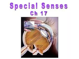
Chapter 8: Special Senses. Sensory Receptors. The sensory division of the peripheral nervous system collects information about sensory stimuli in and around the body. This information is sent to the brain, which generates a motor response to the stimuli.

E N D
Sensory Receptors • The sensory division of the peripheral nervous system collects information about sensory stimuli in and around the body. • This information is sent to the brain, which generates a motor response to the stimuli. • Although sensory receptors are located throughout the body, they are most concentrated in the sense organs—the eyes, ears, nose, mouth, and skin. • Have large, complex sensory organs (eyes/ears) or clusters of receptors (taste buds). • Sensory receptors detect all forms of energy, such as heat, light, pressure, and chemicals.
Vision • Our eyes allow us to see in color and to distinguish fine details and movement. • Your eyes sees lightly different images. Information from both eyes is combined to produce three-dimensional vision. This process is very complex; as much as 30 % of the cerebral cortex processes visual information! • 70% of all sensory receptors in the body are in the eyes! • The adult eye is ~2.5cm in diameter.
Visual Pathway • Light enters the eye through the pupil and passes through the lens, a thick, transparent disk that focuses light on the retina. • The retina is a layer of specialized cells that lines the inner surface of the eye. (more later) • Images are inverted and reversed; brain learns to correct.
Eye Structures • The eye is a hollow sphere; it’s wall composed of three layers: • Fibrous- outermost layer; contains the cornea- transparent coat over the iris, allows light to pass and the sclera- white of the eye- provides shape and protection. • Vascular- middle layer; contains the choroid- lines the sclera, highly vascular to nourish the retina, ciliary body- smooth muscle that alters the shape of the lens, and iris- colored portion of the eye, smooth muscle that controls the amount of light.
Structures cont. • Sensory Layer- Retina- lines the posterior ¾ of the eyeball, contains two types of photoreceptors • Central fovea- cones are most dense, rods are absent, highest visual acuity (resolution). • Optic disc- where the optic nerve attaches, no rods or cones- blind spot.
Retina • The retina contains two types of photoreceptors— rods and cones. • Rodsrespond best to dim light (shades of gray). • Conesrespond best to bright light and are stimulated by specific colors of light (red, green, blue). • ~6 million cones and ~120 million rods • The retina also contains many other neurons that process visual information. The axons of some of these cells make up the optic nerve. • The optic nerve exits through the back of the eye, runs along the base of the brain to the thalamus, and later runs to the cortex for processing.
Accessory Structures • Six external eye muscles- allow eye to follow moving objects. • Eyelidsand eyelashes for protection • Conjuctiva- delicate membrane that secretes mucus • Lacrimal apparatus- tears, contains lysozyme to kill bacteria
Additional Structures • Lens- transparent, focuses light onto the retina, divides the eye into 2 chambers • Pupil- hole in the iris, through which light enters • Anterior segment- contains the aqueous humor- supplies nutrients and oxygen to the lens and cornea • Posterior (vitreous) segment- contains vitreous body- gel-like, keeps shape, keeps retina attached to choroid.
Optical Illusions • http://www.stumbleupon.com/su/1TrSFQ/:p_vcP7@5:6pCyIrKN/jvsc.jst.go.jp/find/mindlab/english/index.html/ • http://colorvisiontesting.com/ishihara.htm#return%20to%20the%20top