Download
1 / 35
350 likes | 355 Views
medically induced CSF leakage in pituitary adenomas. Dr. Rhim Zahedi.

E N D
medically induced CSF leakage in pituitary adenomas Dr. RhimZahedi
CSF rhinorrhea can be easily differentiated from allergic rhinitis or other nasal discharge conditions by biochemical analysis of fluid for sugar (usually more than 30 mg/dL) and beta-2 transferrin, which is present in CSF-containing fluid
Twenty-nine articles were identified from between the years 1980 and 2011, describing 52 patients with spontaneous or medically induced CSF leaks in the setting of a pituitary adenoma
The etiologies of spontaneous CSF leaks are numerous, and leaks have been associated with various factors, including obesity, trauma, and multiparity • There have also been several reports of development of CSF rhinorrhea following the initiation of medical treatment for pituitary adenomas • The classical description of this phenomenon has been in the setting of DA therapy for prolactin-secreting tumors
Results • A majority of these cases (38 patients, 73%) occurred following initiation of medical therapy • whereas the CSF leak developed as the presenting symptom of a pituitary adenoma in the remaining 14 patients (27%) • Forty-two patients (81%) had a prolactinoma • The remaining 10 patients had the following tumor subtypes: nonfunctioning pituitary adenoma (6 patients, 11%), mammosomatotroph cell adenoma (1 patient, 2%), GH-secreting adenoma (2 patients, 4%), and ACTH-secreting adenoma (1 patient, 2%)
36of the 38 patients who developed CSF leaks following initiation of medical therapy had prolactinomas • whereas patients who developed spontaneous (noniatrogenic) CSF leaks had a variety of pituitary adenomas (6 prolactinomas, 6 nonfunctioning pituitary adenomas, 1 GH-secreting adenoma, and 1 ACTH-secreting adenoma) • The medical agents most closely associated with CSF leakage were DA medications in 37 patients (97%) and a somatostatin analog (lanreotide) in 1 patient (3%)
Of the patients who developed CSF rhinorrhea while taking DA medications, 24 were taking bromocriptine and 13 were taking cabergoline • The average time from initiation of medical therapy to onset of rhinorrhea was 3.3 months (range 3 days–17 months) • Seven patients (14%) presented with meningitis in conjunction with CSF rhinorrhea • Tumor size was reported in 9 of 52 cases (17%), with a mean maximum tumor diameter of 3.6 cm
. Forty-nine of 52 patients (94%) were reported to have tumors with neuroimaging evidence of extrasellar tumor invasion • Specific invasion into the sphenoid sinus, ethmoid sinus, or clivus was documented in 29 patients (56%) • In 47 patients, a discrete skull base defect could be identified on imaging studies, most frequently with thin-slice CT images in the coronal plane • The average initial prolactin level in patients with spontaneous CSF leakage was 9169 ng/ ml, compared with 4917 ng/ml in those with medically induced leakage
Treatment Characteristics in Patients With CSF Leaks and Underlying Pituitary Adenomas • In 4 patients, nonoperative management was successfully employed. Of these, 1 patient was successfully treated with temporary lumbar drainage and the other 3 with bed rest or withdrawal of medications • Forty-six patients (88%) ultimately underwent surgical intervention as the definitive treatment for the CSF leak • Definitive procedures for the CSF leaks included: transsphenoidal surgery (32 patients), craniotomy (5 patients), lumboperitoneal shunt (2 patients), and unknown approach (7 patients). • In 21 of 38 cases of DA-induced leak, there was no documented recurrence after the initial treatment
Recurrence was documented in the remaining 17 cases, and a combination of treatment approaches was employed • In 8 cases, temporary cessation of rhinorrhea occurred with treatment reduction or withdrawal, but the rhinorrhea recurred within days or weeks of restarting medical treatment • In 7 cases, rhinorrhearecurred despite initial surgical treatment • Of these cases, rhinorrhea was ultimately resolved with a transsphenoidal approach in 3 patients, a transfrontal approach in 1 patient, lumboperitoneal shunt placement in 1 patient, and craniotomy in 1 patient, and it subsided without treatment in 1 patient
The majority of cases of nonsurgical CSF leaks, the leaks occur in the setting of invasive pituitary tumors (typically prolactinomas) following initiation of standard medical management • In larger and more invasive pituitary adenomas, tumor expansion into the surrounding dural and bony structures is commonly observed • For instance, GH-secreting adenomas and prolactinomas are known to frequently invade the sellar floor and infrasellar space (sphenoid sinus and clivus)
whereas nonfunctioning macroadenomashave a tendency to invade the suprasellarspace via bowing or invasion of the diaphragmasellae • In cases where the arachnoid and/or brain parenchyma have also been violated, the potential for developing a CSF fistula has been established • These communications have little clinical significance as long as the tumor occludes the opening and serves as a “plug,” thus preventing the escape of CSF
Any significant reduction in tumor size, however, can provide a conduit for the escape of CSF, typically resulting in CSF rhinorrhea • Rapid tumor shrinkage is frequently associated with initiation of dopamine agonist therapy for prolactin-secreting tumors • The development of meningitis should be anticipated and addressed in advance in cases of nonsurgical CSF leak. • Surgical repair is the recommended initial treatment for definitive management of DA-induced rhinorrhea, and was ultimately required in nearly 90% of patients reviewed in the literature
Our preferred operation in the majority of cases is the endoscopic endonasal approach, although open surgical repair via a craniotomy may be warranted in a minority of cases • Depending on the tumor subtype and growth patterns, tumor resection may be concurrently achieved prior to reconstruction of the skull base in these instances, although complete tumor resection may be limited in many of these cases as these tumors are by definition invasive into bone and dura • For smaller “weeping” CSF leaks, dural substitutes and/or fibrin glue may be used to achieve a successful repair • For larger CSF leaks, the use of autologous fat or fascia is recommended, with or without a sellar floor buttress • For extremely large or refractory CSF leaks, rotation of a pediclednasoseptal flap may be required to definitively address the CSF fistula
reduction or discontinuation of medical therapy was attempted as the primary treatment in 24% of cases, we do not recommend this strategy in patients who are candidates for surgical repair • The half-life of cabergoline is 63–69 hours and that of bromocriptine is 12–14 hours, suggesting that no immediate effects from medication discontinuation will be achieved via this strategy
We recommend surgical repair of spontaneous or medically induced CSF leaks in the setting of pituitary adenomas, along with safe maximal tumor resection, in patients who are surgical candidates • In patients who are not surgical candidates, a more conservative approach would include temporary cessation of medical therapy and temporary insertion of a lumbar drain, but this strategy is not recommended as a first-line therapy unless deemed medically necessary
A 35-year-old obese male presented with weight gain of over 100 pounds within the last year and absent morning tumescence. Laboratory evaluation was notable for elevated total prolactin (PRL) (4,655 ng/mL) and low total testosterone (24 ng/dL
MRI revealed a large pituitary adenoma (4.2 × 2.5 × 3.5 cm) extending into the cavernous and sphenoid sinuses, partially eroding the sphenoid bone
cabergoline 250 µg twice a week was started. After two doses of cabergoline, the patient developed CSF rhinorrhea • Serum mono total PRL of 370 ng/mL. MRI pituitary revealed persistent tumor, size 4.2 × 3.5 × 2.5 cm. Cabergoline dose was increased to 500 µg twice/week
CSF rhinorrhea completely resolved. Concurrently, monomeric PRL decreased to 90 ng/mL, with a total PRL of 102 ng/mL.
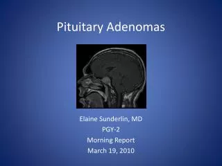
MRI is the modality of choice to provide multiplanar high-contrast images of the pituitary gland and its adjacent structures • (CT) is used only for supplementary purposes, i.e., to look for bony changes or to exclude or visualize calcifications
A standard protocol for MRI of the pituitary and parasellar region consists of thin-section (2–3 mm) sagittal and coronal T1-weighted images with and without contrast enhancement
They are relatively common incidental findings at autopsy (up to 30%) usually remaining asymptomatic
It is thought that arachnoid cysts arise from herniation of an arachnoiddiverticulum through an incomplete diaphragmasellae
typically seen as a small nodule with characteristic high signal at the median eminence in the floor of the third ventricle
Cavernous sinus invasion often restricts complete surgical tumor resection
Intratumoral hemorrhage of macroadenomasoccurs in up to 10–15% of incidental pituitary adenomas
Microadenomas are best seen on coronal images and usually appear hypo- or isointense relative to normal pituitary tissue on unenhanced T1-weighted images
if residual tumor is suspected after incomplete resection of invasive pituitary adenomas. Also in case of persisting or recurring hormone disturbances, MRI is the modality of choice to visualize residual tumor
Due to an en plaque growth pattern often associated with a “dural tail”, differentiation from pituitary macroadenomas usually is not a problem