Download
1 / 1
10 likes | 112 Views
Assessment of [ 3 H] Diprenorphine (DPN) and [ 11 C] Carfentanil (CFN) to Endogenous Opioid Peptide (EOP) Release Darren R. Quelch 1 , Christine A. Parker 1,2 , Salvatore Lanzarone 3 , Vittorio De Santis 3,4 , David J. Nutt 1 , Robin J. Tyacke 1

E N D
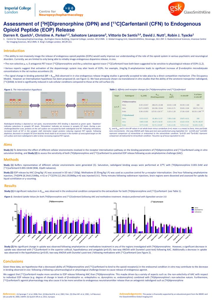
Assessment of [3H]Diprenorphine (DPN) and [11C]Carfentanil (CFN) to Endogenous Opioid Peptide (EOP) Release • Darren R. Quelch1, Christine A. Parker1,2,Salvatore Lanzarone3, Vittorio De Santis3,4, David J. Nutt1, Robin J. Tyacke1 • Centre for Neuropsychopharmacology , Burlington Danes Building, Imperial College, London, W12 0NN. 2. Global Imaging Unit, GlaxoSmithKline, Stevenage, SG1 2NY. 3. Radiochemical Sciences, Imanova Centre for Imaging Sciences, W12 0NN. 4. King’s College London, WC2R 2LS. • Introduction • The ability to non-invasively image the release of endogenous opioid peptides (EOPs) would vastly improve our understanding of the role of the opioid system in various psychiatric and neurological disorders. Currently, we are limited to only being able to reliably image endogenous dopamine release, in vivo. • The non-selective µ, κ, δ antagonist PET tracer [11C]Diprenorphine and the µ selective agonist tracer [11C]Carfentanil have both been suggested to be sensitive to physiological release of EOPs (1,2). • Various reports suggest that activation of the dopaminergic system may alter levels of EOPs. For example, 2mg/kg D-amphetamine leads to significant increase of β-endorphin microdialysate concentrations in the rat nucleus accumbens (3). • The signal change in binding potential (BP = Bmax/Kd) observed in in vivo endogenous release imaging studies is generally accepted to take place by a direct competition mechanism (The Occupancy Model). However an Internalisation hypothesis has been proposed (4; see Figure 1). We have previously shown via translational in vitro studies that the ability of the serotonin transporter radioligand, [11C]DASB, to bind is significantly reduced in sub-cellular conditions compared to those at the cell surface (5). Table 1. Affinity and receptor changes for [3H]diprenorphine and [11C]carfentanil Figure 1. The Internalisation Hypothesis Radioligand binding is depicted as red spots, neurotransmitter (NT) binding is depicted as green spots. ‘Depletion’ indicates reductions in synaptic NT levels. ‘Stimulation’ indicates an increase in synaptic NT levels. During a pre-challenge/baseline scan, proteins at the cell surface are occupied by both radioligand and NT. Following stimulation, increased levels of NT in the synaptic cleft internalise target proteins reducing regional PET signals. Following depletion, decreases in synaptic NT from baseline levels leads to an increase in the number of unoccupied target on the cell surface and more radioligand is able to bind, increasing regional PET signals. Kd and Bmax and in vitro BP values in rat whole brain minus cerebellum (n=4, mean ± s.e.mean) in Extra, Intra and Endo ionic environments. One way-ANOVA with Tukey post-test were performed using SigmaStat 3.0. *p<0.05 and **p<0.001 represent comparison of intracellular or endosomal to the extracellular condition. #p<0.05 and ##p<0.001 represent comparison of endosomal to intracellular condition. $Assumes 1g of tissue equivalent to 1ml. Aims Study (1) To determine the effect of different cellular environments involved in the receptor internalisation pathway on the binding parameters of [3H]diprenorphine and [11C]carfentanil using in vitro saturation binding, and Study (2) to assess the sensitivity of both [3H]diprenorphine and [11C]carfentanil to potential EOP release following acute amphetamine challenge (AAC). Methods Study (1) Buffers representative of different cellular environments were generated (5). Saturation, radioligand binding assays were performed at 37⁰C with [3H]diprenorphine 0.001-3nM and [11C]carfentanil 0.003-10nM (NSB, 10µM naloxone). Study (2) EOP release by AAC (2mg/kg I.P.) was assessed in SD rats (~250g). Methadone (0.35mg/kg I.P.) was used as a positive control for μ-receptor internalisation. One hour following amphetamine injection, [3H]DPN (8.24±0.21MBq, n=5) or [11C]CFN (12.24±1.053MBq,n=4) was injected (I.V.). Thirty minutes following radiotracer injections, brain regions were dissected and assessed for uptake by liquid scintillation or γ-counting. Results Study (1) A significant reduction in Bmax was observed in the endosomal condition compared to the extracellular for both [3H]diprenorphine and [11C]carfentanil(see Table 1). Figure 2. Standard Uptake Values for both [3H]Diprenorphine and [11C]Carfentanil following AAC and methadone treatment. Analysis performed with SigmaStat version 3.0. Study (2) No significant change in uptake was observed following amphetamine or methadone treatment in any of the regions investigated with [3H]diprenorphine. However, asignificant decrease in uptake was observed with [11C]carfentanil in the superior colliculi, hypothalamus and amygdala (p<0.05; two-way ANOVA with Dunette’s post-test) following AAC. Additionally a decrease in uptake was observed in the hypothalamus (p<0.05; two-way ANOVA with Dunette’s post-test ) following methadone with [11C]carfentanil (see Figure 2). Conclusions Given these data, we hypothesise that a decreased ability of [3H]diprenorphine and [11C]carfentanil to bind to the opioid receptor(s) in the endosomal condition in vitro may contribute to the decrease in binding observed in vivo following a following a pharmacological or physiological challenge known to cause release of endogenous agonist. We suggest that [11C]carfentanil maybe more sensitive to EOP release following AAC than [3H]diprenorphine. This maybe driven by a variety of aspects such as: the non-selectivity of AAC with respect to individual EOP release (e.g. degree of change in levels of β-endorphin vs. enkephalins vs. dynorphins vs. endomorphins) combined with [3H]diprenorphine’s non-selective nature. Furthermore, [11C]carfentanil’s agonist pharmacology may also cause it to be more sensitive to endogenous neurotransmitter release than an antagonist radioligand such as [3H]diprenorphine Acknowledgements: This poster is financially supported by an educational grant from the BBSRC and the GlaxoSmithKline Global Imaging Unit References: (1) Sprenger T. et al, 2006, Pain. (2) Bencherif B. et al, 2002, Pain. (3) Olive MF. et al, 2001, J of Neurosci. (4) Laruelle M, 2000, JCBFM. (5) Quelch DR et al, 2012, Synapse.