Download
1 / 38
380 likes | 389 Views
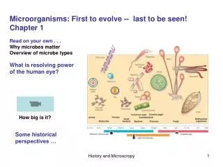
Just what are microbes made of?. Lecture Chapter 4 Prokaryotic and eukaryotic cells Prokaryotic cell features Chapter 3 (next class) Endosymbiotic theory Light microscopy Electron microscopy Microscopy techniques and staining. Lab Aseptic technique Microbes in the environment

E N D
Just what are microbes made of? Lecture Chapter 4 Prokaryotic and eukaryotic cells Prokaryotic cell features Chapter 3 (next class) Endosymbiotic theory Light microscopy Electron microscopy Microscopy techniques and staining Lab Aseptic technique Microbes in the environment Oil immersion microscopy Pre-labs Pure culture Motility
CHAPTER 4 Prokaryotic vs. Eukaryotic cells
Odd bacterial cell shapes Figure 4.5 - Overview
Bacterial cell arrangements Figure 4.1 - Overview
Prokaryotic cell overview Figure 4.1 - Overview
Prokaryotic cell features • Glycocalyx
Prokaryotic cell features • Glycocalyx • Flagella Figure 4.7 - Overview
Prokaryotic cell features • Glycocalyx • Flagella • Axial filaments (endoflagella) Spirochete Leptospira interrogans Figure 4.7 - Overview
Prokaryotic cell features • Glycocalyx • Flagella • Axial filaments (endoflagella) • Attachment pili (fimbriae)
Prokaryotic cell features • Glycocalyx • Flagella • Axial filaments (endoflagella) • Attachment pili (fimbriae) • Conjugation pili (sex pili)
Prokaryotic cell features • Glycocalyx • Flagella • Axial filaments (endoflagella) • Attachment pili (fimbriae) • Conjugation pili (sex pili) 6. Cell wall Figure 4.13 - Overview
Independent Learning 1. Review cell wall structure in bacteria. You should know the structure inside and out. Literally.
Prokaryotic cell features 7. Plasma membrane Figure 4.14 - Overview
Plasma membrane: osmosis and tonicity Figure 4.18 - Overview
Prokaryotic cell features 8. Ribosomes ProkaryoticEukaryotic 3 RNAs (23s, 16s, 5s) 4 RNAs (28s, 15s, 5.8s, 5s) 53 proteins 70 proteins 30S/ 50S subunits 40S/ 60S subunits 70S ribosome 80S ribosome Figure 4.19
Prokaryotic cell features 9. Endospores
CHAPTER 3 Microscopy
Compound light microscope Figure 3.1a
Refraction and immersion oil Figure 3.3
Brightfield and darkfield microscopy Figure 3.4 - Overview
Phase contrast and Nomarski optics (DIC) Figure 3.5 Figure 3.4 - Overview
Fluorescence and confocal microscopy Figure 3.6 Figure 3.7
Transmission Electron Microscopy (TEM) Figure 3.9a
Scanning Electron Microscopy (SEM) Figure 3.9b
SEM images Didinium eating Paramecium (protozoa) Protozoan Radiolarian Fungus Aspergillus
SEM images Bacillus anthracis sporulation (bacterium) Alga Ceratium Penicillium notatum conidiophore (fungus) SEMs courtesy of Dennis Kunkel Inc.
Independent study • Look at the evidence for the endosymbiotic theory. Be prepared to • present the evidence that mitochondria and chloroplasts arose from a • symbiotic interaction between an early eukaryote and a prokaryote. • Review aerobic respiration (see figure 5.17). • Review the light dependent and light independent reactions of • photosynthesis (see Figure 5.24 and 5.25).
Microscopy Basics • Living preparations
Microscopy Basics Living preparations Stained preparations
Differential stains- The Gram Stain Figure 3.11a
Differential and special stains Figure 3.12 and 3.13