Download
1 / 40
410 likes | 580 Views
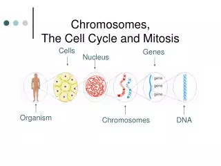
Chromosomes and Cell Cycle. Cell Division Cell Cycle Mitosis Cytokinesis. Cell Basics. There are trillions of cells in your body. Cells are microscopic Cells have DNA inside a structure called the nucleus The nucleus is “surrounded” by a structure called the nuclear envelope

E N D
Chromosomes and Cell Cycle • Cell Division • Cell Cycle • Mitosis • Cytokinesis
Cell Basics • There are trillions of cells in your body. • Cells are microscopic • Cells have DNA inside a structure called the nucleus • The nucleus is “surrounded” by a structure called the nuclear envelope • Cells are filled with a jelly like substance called the cytoplasm
A Cell Nuclear Envelope Nucleus DNA Cytoplasm
Cell Division • Cell division is the splitting of a single cell into two daughter cells that are identical to each other • mitosis– process of nuclear division (dividing the nucleus) • cytokinesis- process of division of the cytoplasm
Cell Division Cells divide because the organism needs to: • Grow- our cells don’t get bigger in size, they get bigger in number • Repair- needed because of worn out or injured cells (your skin cells are replaced every 28 days; your stomach every 7) • Reproduce: • asexual – one parent; offspring identical to parent – mitosis or binary fission • sexual – combination of genetic material from two parents – meiosis (more on this later!)
Asexual • asexual reproduction • binary fission
Asexual • asexual reproduction • mitosis
The Cell Cycle Cell Cycle- • occurs in somatic cells • What are somatic cells? • a set of events that results in two new daughter cells, which then start the process again. • Interphase • G1 S G2 • Prophase • Metaphase • Anaphase • Telophase • Cytokinesis Growth and Preparation- 90% of the time spent here Mitosis- division of the nucleus Division of the cytoplasm
The Cell Cycle Interphase • 90% of the time, the cell is in this phase • the cell grows • performs operations unique to the type of cell (stomach cells make digestive enzymes, some white blood cells make antibodies, etc) Three stages of interphase: • G1 (growth stage 1) • S (synthesis) • G2 (growth stage 2)
The Cell Cycle • GROWTH 1 STAGE – G1 • decides whether or not the cell will divide • makes its structural proteins and enzymes to perform its functions • a pancreas cell will produce and secrete insulin • salivary gland cells will produce and secrete enzymes in the mouth to aid in digestion
The Cell Cycle S Synthesis (DNAReplication) • each of the chromosomes is copied
The Cell Cycle GROWTH 2 PHASE – G2 • DNA replication is checked by DNA repair enzymes • cell prepares for mitosis • proteins organize themselves to form a series of fibers called the spindle
DNA correctly replicated? All proteins built for cell division?
INTERPHASE IN AN ANIMAL CELL INTERPHASE IN A PLANT CELL Note that the DNA is in the form of chromatin – loose and in long strands. The nucleolus is usually visible during interphase, but not during mitosis
Mitosis • Follows interphase when the cell is ready to divide • 4 main parts • prophase, metaphase, anaphase, telophase • P-MAT
PROPHASE ANAPHASE METAPHASE TELOPHASE
MITOSIS • PROPHASE • condensing of 2 sister chromatids • chromatin coils up • chromosomes become visible • centriolesreplicate and begin to move to opposite sides of the cell • nuclear envelope (the outside of the nucleus) disappears
Notice that the chromatin begins to coil up and you see “space” in the nucleus between what will soon be evident as separate chromosomes
MITOSIS • METAPHASE • spindle fibers move the chromosomes to the middle • this organization helps to ensure that in the next phase, when the chromosomes are separated, each new nucleus will receive one copy of each chromosome
Chromosomes in metaphase have their centromeres lined up in the middle and their long arms are trailing from each side. Some students think it looks like Chinese characters, others have compared it to stitches on a zombie mouth.
MITOSIS • ANAPHASE • each chromosome is attached to a spindle which moves it toward one pole • chromatidsmove apart from one another • results in equal separation and distribution of chromosomes
In anaphase the centromeres are in rows at each end of the cell and the arms of the chromosomes are trailing away toward the middle of the cell Some students think this looks like a scary zombie mouth opening
MITOSIS • TELOPHASE • newly separated chromatidsarrive at opposite ends of cell • nuclear envelope reappears around the daughter nuclei • the chromosomes uncoil and are no longer visible • cytokinesismay also begin during this stage • this phase is opposite of prophase in the events that happen • end with two new nuclei (one for each new cell)
In telophase the 2 new cells are preparing to enter interphase The chromosomes uncoil In this picture you can see the cell wall forming between the two “wads” of chromatin This is called a cell plate until it reaches both sides of the old cell and divides it into 2 new cells
CYTOKINESIS • Process in which the cytoplasm divides and two separate cells (daughter cells) form • In animals, it begins with the formation of a cleavage furrow • In plants, a cell wall forms
Plant cell plate Animal cell cleavage
Challenge! • What differences do you see between animal cell division and plant cell division? (Hint: does one type of cell have something the other doesn’t?) 1. 2. 3.
Challenge! • Shape • animal cells are round • plant cells are square • Structures • animal cells have structures called centrioles • plant cells do not • Telophase • a cleavage furrow divides animal cells • a cell plate divides plant cells
MITOSIS—Summary PROPHASE- chromosomes visible (P for Phat), nuclear membrane disappearing chromosomes condense/fatten and becomevisible) METAPHASE- sister chromatids lined up in the middle/equator (M for middle, chromosomes lined up in the middle of cell)
ANAPHASE- sister chromatids pulled apart (A for Apart or Away because the chromatids pull apart and move away from center) TELOPHASE- chromosomes are at ends of cell, cells prepare to separate (T for Two new nuclear envelopes are forming) Cleavage furrow Cell plate
Not all cells reproduce… • some leave the cell cycle here and do not undergo cell division • red blood cells – which “kick out” their nucleus to make room for the hemoglobin and therefore can’t divide • brain and spinal cord cells – rarely if ever divide; called G0 (pronounced G naught)
Abnormal Cell Cycle: Cancer What causes cancer? • Cancer is caused by mutations (changes) in the DNA, including the genes that regulate the cell cycle • Basically: • uncontrolled cell growth • cancer cells grow and divide as long as they receive nutrients • cancer cells crowd normal cells causing tissues and organs to stop working
Environmental factors can increase the risk of cancer. • Substances that are known to cause cancer are called carcinogens(kar SIH nuhjunz). • Tobacco, tobacco smoke, and alcohol, are examples of carcinogens • Some viruses are linked to cancer • HPV (human papilloma virus) can lead to cervical cancer as well as other forms of cancer • Mutagens cause mutations, which can lead to cancer. • Radiation (x-rays, UV light) is a mutagen. So are some chemicals.