Download
1 / 1
10 likes | 131 Views
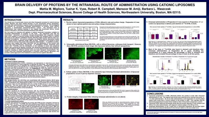
BRAIN DELIVERY OF PROTEINS BY THE INTRANASAL ROUTE OF ADMINISTRATION USING CATIONIC LIPOSOMES Mattia M. Migliore, Tushar K. Vyas, Robert B. Campbell, Mansoor M. Amiji, Barbara L. Waszczak

E N D
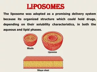
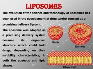
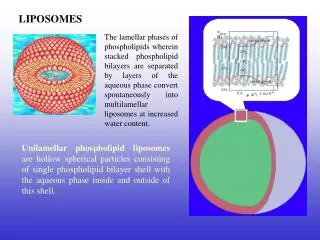
BRAIN DELIVERY OF PROTEINS BY THE INTRANASAL ROUTE OF ADMINISTRATION USING CATIONIC LIPOSOMES Mattia M. Migliore, Tushar K. Vyas, Robert B. Campbell, Mansoor M. Amiji, Barbara L. Waszczak Dept. Pharmaceutical Sciences,Bouvé College of Health Sciences, Northeastern University, Boston, MA 02115. INTRODUCTION The clinical use of neurotrophic factors and other therapeutic proteins for central nervous system diseases has been limited because of poor blood-brain barrier penetration. Currently, their administration requires invasive intracerebral infusions. We investigated the potential for non-invasive intranasal administration of proteins using cationic liposomes, a phospholipid nanoparticle-delivery system. The intranasal route of administration was selected because it can bypass the blood-brain barrier, avoids systemic absorption, and limits potential peripheral sideeffects. Our first goal is to develop and optimize a cationic liposomal formulation suitable for intranasal administration of proteins to the brain. Our hypothesis was that the electrostatic interaction of positively charged phospholipids with negatively charged sialic acid residues of mucous proteins would increase the residence time of cationic liposomes at the olfactory epithelium thereby affording higher protein delivery than non-liposomal formulations. As proof or principle, we prepared cationic liposomes loaded with a model protein, ovalbumin (OVAL; MW= 45 KDa). Transport of the protein to brain was studied at various time points after intranasal administration. In qualitative studies, liposomes were loaded with Alexa 488-OVAL and the distribution of the protein was determined using fluorescence microscopy. In quantitative studies, liposomes were loaded with 111In-OVAL and the distribution of the radiolabel to brain and peripheral tissues was monitored by gamma counting. Two liposomal preparations were evaluated: OVAL was added to either preformed liposomes, or to the aqueous phase during liposome formation resulting in different particle sizes and brain transport characteristics. The results show that intranasal administration of proteins using cationic liposomes may provide a useful approach to the treatment of central nervous system disorders. . RESULTS 5. Intranasal administration of Preparation # 2 was superior to Preparation # 1 or control (no liposomes) in delivery and retention of 111In-OVAL in brain. 1. The two cationic liposomal preparations of OVAL differed in size and surface charge. Preparation # 2 was shown to have optimal particle size and surface charge characteristics. Time course of brain uptake of 111In-OVAL (1 µg/ µl) from liposomal preparations or control (PBS) Preparation # 2 had significantly lower particle sizes than Preparation #1. A smaller particle size may result in better paracellular transport to brain by the intranasal route. Preparation #1 and #2 (1 g/l) had similar surface charges, both within the optimal range for liposomal adherence to tissue. The more concentrated (2 g/l) form of Preparation #2 had a significantly higher surface charge than the 1 g/l formulations. Higher surface charge might be predicted to prolong tissue residence time and result in higher protein transport to brain. The protein loading efficiencies for both preparations were similar and exceeded 90%. This indicates that both preparations incorporate nearly all of the added protein, and that the amount of protein delivered in equal volumes of each preparation should be equivalent (~50 g of OVAL). Data presented as mean ± SEM. Brain levels as % of administered dose / g tissue at 1 hr, 4 hrs, 6 hrs, and 24 hrs after intranasal administration (mean ± SEM). The highest brain levels occurred at 1 hr after intranasal administration of both Preparation #2 and control (no liposomes). However, by 4 hrs and 24 hrs after administration, brain levels were significantly higher for Preparation #2 than for Preparation #1 or control (P= 0.0468, unpaired Student’s t-test, and P<0.01,one way ANOVA and Tukey’s post hoc test, respectively). This suggests that liposomal Preparation # 2 results in longer retention of the protein in brain. 2. Intranasally administered Alexa 488-OVAL, with or without liposomes, undergoes brain transport. However, Alexa 488-OVAL persists in brain longer after liposomal delivery than without nanoparticles. 6. Most of the dose of 111In-OVAL was found in stomach and intestines after intranasal administration of Preparation #2 (1 μg/μl). Doubling the concentration to 2 μg/μl reduced the percentage of dose in stomach and intestines and increased the percentage of dose in brain at all time points. Biodistribution studies of intranasal 111In-OVAL in Preparation # 2 (1 μg/μl and 2 μg/μl) METHODS Preparation of Cationic Liposomes: Dioleoylphosphatidylcholine (DOPC), cholesterol, and stearylamine in chloroform were mixed (40:10:50) and placed in a rotary evaporator to eliminate the chloroform. The resulting lipid film was freeze-dried for 4 hrs to eliminate any remaining chloroform. Phosphate buffered saline (PBS) was then added to the lipid film at a 1:1 ratio followed by vortexing. The resulting cationic liposomes were then sonicated for 5 min. For Preparation #1, OVAL was added to preformed liposomes (1 µg protein/μl of liposomal solution) and vortexed. For Preparation #2, liposomes were prepared as above except that OVAL was added to the PBS in the hydration step. This aqueous solution of 1 µg protein/µl (or 2 µg protein/µl for the concentrated formulation), was added to the lipid film, vortexed, and sonicated for 10 min. Following sonication, liposomes were centrifuged for 5 min to separate the liposomes from the un-encapsulated protein. The top layer, containing the liposomes with the encapsulated protein, was collected and characterized using a Brookhaven Instruments particle size and zeta potential analyzer. Intranasal administration: Male Sprague-Dawley rats (225-300g) were anesthetized with ketamine/xylazine (80/20 mg/kg respectively), and were placed in a supine position with their heads at an angle of administration of 90°. Formulations were administered at either 5 µl or 2.5 µl increments (for the concentrated formulation) every 2-4 min alternating nares for a total of either 25 µl or 12.5 µl per nostril respectively. The total protein dose was nominally 50 µg/rat. Following administration, the animals remained in a supine position for 60 min. The rats were sacrificed at 1, 4, 6 or 24 hrs after intranasal administration of the formulations. Immunohistochemistry: Rats were sacrificed by transcardial perfusion with 4% paraformaldehyde. Brains were collected, post-fixed for 2 hrs, and then submerged in 30% sucrose for 48-72 hrs. Cryostat sections, 25 µm thick, were collected from different areas of the brain, including the substantia nigra (SN) and striatum. Immunohistochemistry for tyrosine hydroxylase was performed using a rabbit monoclonal anti-tyrosine hydroxylase antibody (Chemicon) and anti-rabbit secondary antibody coupled to Texas Red (Jackson ImmunoResearch). Immunohistochemistry for OVAL was performed using a polyclonal rabbit anti-OVAL antibody (Chemicon), and anti-rabbit secondary antibody coupled to Texas Red (Jackson ImmunoResearch). 111In-OVAL biodistribution studies: 111In-OVAL was prepared by conjugating the protein with DTPA succinic anhydride, followed by chelation with 111In ions. 111In-OVAL was then purified by gel filtration and dialyzed to remove free 111In. The labeled protein was then incorporated into both cationic liposomal preparations as previously described. 111In-OVAL in PBS (no nanoparticles) was used as a control. Male rats (225g-300g) were intranasally administered a total of 50 µl (~1µg/µl) of radiolabeled protein using the protocol above. Initially, these experiments were conducted using both preparation #1 and #2 (with sacrifice at 6 hrs and 24 hr time points). In subsequent experiments, only preparation # 2 was studied (at 1hr, 4 hr, 6 hr, and 24 hr time points) since preliminary data showed that preparation # 2 resulted in higher protein transport to brain. 111In OVAL in Preparation #2 (2 µg/µl) 111In OVAL in Preparation #2 (1 µg/µl) Intranasal Alexa 488-OVAL (Preparation # 2) 24 hr time point, SN (20x) Intranasal Alexa 488-OVAL (Preparation # 1) 24 hr time point, striatum (20x) Intranasal Alexa 488- OVAL (Preparation # 2) 6 hr time point, SN (20x) Intranasal Alexa 488-OVAL (no nanoparticles) 24 hr time point, striatum (20x) Intranasally administered Alexa 488-OVAL (no nanoparticles) is detectable in brain by 6 hrs. Somewhat lower levels remain at 24 hrs (left panel). Both liposomal preparations also deliver the protein to brain after intranasal administration. Note the deposits of punctate fluorescence in the striatum and SN at 6 and 24 hrs (middle and right panels). In SN, there is apparent cellular uptake of the protein by 24 hr after administration of the liposomal preparations. Cellular uptake is not seen in the SN at the 6 hr time point (right panels), nor after administration of Alexa 488-OVAL without nanoparticles (data not shown). 3. Cellular uptake of Alexa 488-OVAL in the substantia nigra following intranasal administration of liposomal preparations occurs in part by dopamine neurons. Time course of brain uptake of 111In-OVAL (2 µg/µl) from liposomal Preparation # 2 or control (PBS) Intranasal Alexa 488-OVAL (Preparation # 2) 24 hr time point, SN (40x) TH positive dopamine neurons SN (40x) Merged image Brain levels as % of administered dose / g tissue at 1 hr, 4 hrs, 6 hrs, and 24 hrs after intranasal administration (mean ± SEM).111In-OVAL( 2 µg/ µl) in liposomal Preparation #2 resulted in significantly higher brain levels when compared to control at all time points (P= 0.0227, 0.0005, 0.0390, 0.0387 respectively; unpaired Student’s t-test). Intranasal Alexa488-OVAL (Preparation # 2) 24 hr time point, SN (40x) Intranasal administration of liposomal Alexa-488 OVAL results in apparent cellular uptake in the SN (left). At least some of the cells taking up the protein stain positive for tyrosine hydroxylase (TH) indicating that they are dopamine neurons (above). • CONCLUSIONS • Liposomal preparations of OVAL effectively deliver the protein to brain after intranasal administration to rats. The highest brain levels were detected at the shortest time point, i.e. 1 hr after administration. By 24 hrs after administration, both Preparation #1 and #2 yielded higher brain levels than a control solution of the protein in PBS. This indicates that the liposomal preparations increase brain residence time of the protein compared to control. • Liposomal OVAL delivered intranasally yields discrete protein deposits in both striatum and SN by 6 hrs after administration, with apparent cellular uptake in the SN by 24 hrs. • Intranasal administration of the 2 µg/µl form of liposomal Preparation #2 provides higher brain levels and reduced distribution to the GI tract relative to the more dilute form. 4. Protein integrity of liposomal OVAL following intranasal administration is not altered. OVAL was detected in the brain by immunohistochemistry following intranasal administration of unlabeled OVAL in cationic liposomes (preparation # 2). Note fluorescent deposits in both SN and striatum. This suggests that fluorescent deposits shown previously with Alexa 488-OVAL are due to the presence of the intact ovalbumin protein, and not to the unassociated Alexa-488 fluorophore. Intranasal OVAL (Preparation # 2, 2 μg/μl) 1 hr time point, striatum (20x). Intranasal OVAL (Preparation # 2, 2 μg/μl) 24 hr time point, SN (40x). This work was supported by a 2005-2006 American Foundation for Pharmaceutical education fellowship, and the NCI-NSF IGERT Nanomedicine S&T award.