Download
1 / 72
780 likes | 1.27k Views
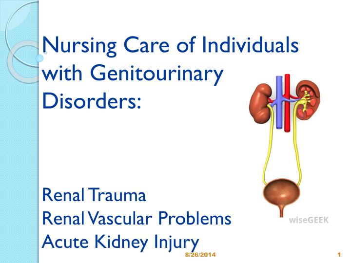
Nursing Care of Individuals with Genitourinary Disorders: Renal Trauma Renal Vascular Problems Acute Kidney Injury. The Kidney. Primary function Regulate volume and composition of ECF (extracellular fluid) Excrete waste products Other functions Regulate acid-base balance Control BP

E N D
Nursing Care of Individualswith Genitourinary Disorders:Renal TraumaRenal Vascular ProblemsAcute Kidney Injury
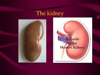
The Kidney • Primary function • Regulate volume and composition of ECF (extracellular fluid) • Excrete waste products • Other functions • Regulate acid-base balance • Control BP • Produce Erthyropoietin • Activate Vitamin D
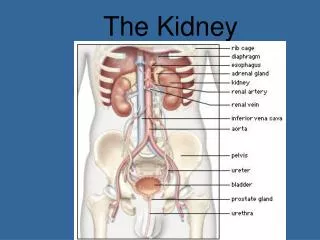
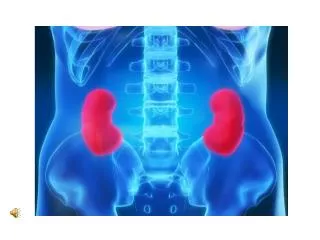
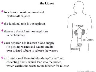
Kidney- macrostructure • kidney anatomy
Kidney- microstructure • nephron
The Nephron • Why is it called the functional unit of the kidney?
Glomerular Filtration Rate • Glomerular filtration rate • Used to assess how well the kidneys are working • Estimates how much blood passes through the glomeruli each minute • The amount of filtrate formed per minute by the two kidneys combined
Glomerular Filtration Rate • For average male GFR is 125ml/min • That would create180 L/d! • More than 99% of the filtrate is reabsorbed • Average 1mL/min of urine excreted • 1-2 L/day • Older people will have lower normal GFR levels, because GFR decreases with age
GFR • GFR too high • increased urine output • threat of dehydration and electrolyte depletion • GFR too low • insufficient excretion of wastes • GFR of 60 or higher is in the normal range • GFR below 60 may mean kidney disease • GFR of 15 or lower may mean kidney failure
The Kidney • Primary function • Regulate volume and composition of ECF (extracellular fluid) • Excrete waste products • Other functions • Regulate acid-base balance • Control BP • Produce Erthyropoietin • Activate Vitamin D
Functions of the Kidneys • Regulates acid-base balance • HCO3 and H+ • Controls Blood Pressure: • Renin Release
RAAS • Kidney senses low perfusion • Renin released by kidney • Angiotensinogen (from liver) acivated into angiotensin I • Converted to Angiotensin II by ACE • Angiotensin II stimulates release of aldosterone • Na+ and H2O retained
Functions of the Kidneys • Erythropoietin Release • If a patient has chronic kidney disease or chronic renal failure, what condition will occur and why?
Functions of the Kidneys • Erythropoietin promotes the formation of RBC’s in response to decreased O2 carrying capacity • Anemia from impaired erythropoietin production and platelet abnormalities > bleeding risk
Functions of the Kidneys • Activated Vitamin D • Necessary to absorb Calcium in the GI tract. There is decrease in synthesis of D3, the active metabolite of Vitamin D If a patient has renal failure, what will happen to the patient’s serum calcium level?
Functions of the Kidneys • Inability of kidneys to activate vitamin D- hypocalcemia > parathyroid gland > secretes PTH > stimulates bone demineralization > release calcium from bones • Low serum calcium level/elevated phosphate • Why do you have a elevated serum phosphate?
Review- Functions of the Kidney • Regulate • Volume & composition of extracellular fluid • F&E balance • Acid/Base balance • Blood pressure regulation • Erythropoetin release • Vitamin D activation
Acute Kidney Injury • Rapid decline in renal function that leads to accumulation of nitrogenous wastes in the blood (azotemia) • Etiology of AKI: • Pre-renal • Intra-renal • Post renal
Acute Kidney InjuryPre-renal Hypovolemia dehydration, shock, burns Decreased cardiac output CHF, MI, arrhythmias Decreased vascular resistance septic shock Renal vascular obstruction renal artery stenosis, thrombus Causes related to decreased blood flow to the kidneys
Acute Kidney InjuryIntra-renal Conditions causing direct damage to renal tissue causing damage to nephrons • Result from ischemia • Nephrotoxins • Hemoglobin released from hemolysis of red blood cells • Myoglobin released from necrotic muscle cells
Acute Kidney InjuryIntra-renal Primary Renal Disease • Acute glomerulonephritis/pyelonephritis • Systemic lupus • Acute Tubular Necrosis (ATN) • Necrosis of tubular cells which slough and plug tubules • Potentially reversible • Most common cause of intra-renal AKI
Acute Tubular Necrosis(ATN) • Renal ischemia • Disruption basement membrane;destruction tubular epithelium • Nephrotoxic agents • Necrosis tubular epithelium… plug tubules; basement membrane intact. • Potentially reversible IF • Basement not destroyed and tubular epithelium regenerates Renal ischemia Nephrotoxic agents
Acute Kidney InjuryIntra-renal • Acute Tubular Necrosis (ATN) • Nephrotoxic drugs/chemicals (ATN) • Aminoglycosides* • Radiographic contrast agents • Arsenic, lead, carbon tetrachloride
Acute Kidney InjuryIntra-renal • Hemolytic blood transfusion (ATN) • Trauma • crushing injuries which release myoglobin • damaged muscle tissue and blocks tubules (rhabdomyolysis)(ATN) • What is Rhabdomyolysis?
Healthy ATN Compare & Contrast
Lupus Nephritis • ‘Flea bite’ look
Acute Kidney InjuryPost-renal • Mechanical obstruction of urinary outflow • urine backs up into renal pelvis • BPH • Calculi • Trauma • Prostate cancer
Stages of Acute Kidney Injury • Initiating Phase • Time of insult until signs and symptoms become apparent • Oliguric Phase • Usually appears 1-7 days of initiating event • Diuretic Phase • Start varies, usually within10-12 days of onset oliguric phase • Recovery • Usually within a month, recovery takes up to 12 months
Urine output in AKI varies widely & does NOT provide clinical correlation to the degree of injury!!!!! • Must look at GRF
Oliguric Phase • Onset- 1-7 days • Duration- 10-14 days • Urine Output- Less than 400 ml/24 hours in 50% of patients (Can have non-oliguric AKI) • Signs & Symptoms to anticipate- • Specific gravity fixed at 1.010 in oliguria in intra renal failure – may be elevated in pre & post • Fluid overload • Urine with RBCs, casts, WBCs, protein (if glomerulus damaged) • K+ likely elevated
Oliguric Phase • Metabolic acidosis • kidneys unable to synthesize HCO3, cannot excrete H+ and acid metabolites, serum bicarbonate decreased because used to buffer H+ • Kussmaul breathing • Calcium deficit & phosphate excess • decreased GI absorption of Ca (Vit D) • increase in Calcium secretion • Nitrogenous product accumulation • unable to eliminate urea and creatinine > elevated BUN, serum creatinine
Treatment – Oliguric phase • Fluid Challenge/Diuretics • Done to r/o dehydration as cause of ARF and to blast out tubules if ATN • 250-500cc NS given I.V. over 15 minutes • Mannitol (osmotic diuretic) 25gm I.V. given • Lasix 80mg I.V. given • Should see what within 1-2 hours?
Treatment – Oliguric phase • If fluid challenge fails- intake limited • Fluid restriction • 600ml + u.o. past 24 hours • Patient’s u.o. yesterday was 300ml. What will be the allowed fluid intake today?
Diuretic Phase • Onset- days to weeks • Duration- 1-3 weeks • Urine Output- 1-3 liters/day • Signs & Symptoms to anticipate • Elevated BUN and Serum Creatinine • What happens to intravascular volume? • What happens to BP? • Urine Na? • K+ elevated or decreased?
Recovery Phase • Onset- When BUN and Creatinine stabilized • Duration- 4-12 months • Urine Output- Normal • Signs & Symptoms • Continue to monitor for signs and symptoms of F & E imbalances • All body systems for effects of fluid volume changes • What are some key nursing interventions?
Diagnostic tests in AKI • BUN (blood urea nitrogen) • Measurement of amount of urea in blood • Normal -6-20 mg/dl • What is urea? • BUN fluctuates • BUN elevated when? • BUN decreased when?
Diagnostic tests in AKI • Serum Creatinine • End product of muscle and protein metabolism • Excreted by the kidneys at a constant rate • Normal = 0.6 – 1.3 mg/dl • Directly related to GFR • 2 X normal (2.4) = 50% nephron fx loss • 10 X normal (12) = 90% nephron fx loss • More accurate indicator of renal function than BUN • BUN:Creatinine ratio Normal= 12:1 to 20:1
Diagnostic tests in AKI • Creatinine clearance • Normal= 120-125ml/minute • Most accurate indicator of Renal Function • Reflects GFR • Involves a 24 hr urine/serum creatinine • Formula: urine creatinine X urine Volume serum creatinine
24 hour urine • What is the nurses role in the collection of a 24 hour urine? • What if they have a foley cath?
Diagnostic tests in AKI • Urine Specific Gravity • Normal= 1.003-1.030 • Will be fixed a 1.010 usually in AKI due to kidneys losing ability to concentrate urine • Serum Electrolytes • Sodium • Potassium • Calcium • Phosphorus
Diagnostic tests in AKI • Serum Electrolytes • Serum Sodium • Normal= 135-145 • May be high, low, or normal • When would it be high/low?
Diagnostic tests in AKI • Serum Electrolytes • Serum Potassium • Normal= 3.5-5 meq/L • Almost always increased in renal failure • Why? Two major reasons • If > 6.0 treatment to prevent….
Diagnostic tests in AKI • Serum Electrolytes • Serum Phosphorus • Normal=2.8-4.5mg/dl • Almost always increased. Why? • What other process is occurring to increase serum phosphorus?
Diagnostic tests in AKI • Serum Electrolytes • Serum Calcium • Normal=9.0-11.0 mg/dl • Almost always decreased, why? • What other process is occurring to decrease serum calcium?
Diagnostic tests in AKI • ABGs • Metabolic acidosis-due to decreased ability of kidneys to excrete acid metabolite (uric acid) • So the pH will be high or low? • Bicarb- decreased due to bicarb being used up to buffer excess H+ ions
Management of AKI • Treat the primary disease/condition • Prevention • Frequent monitoring for early signs of AKI in at risk patients • What are these signs?
Management of AKI • Assess for FVD vs FVE • VS • Strict I&O • Daily weights • Monitor labs- which ones? • Metabolic acidosis • Administer NaHCO3 IV as ordered
Management of AKI • Hyperkalemia • Insulin and glucose • K+ moves back into the cells when insulin is given. • Glucose to prevent hypoglycemia • Sodium Bicarbonate • Correct acidosis and shifts K+ into cells • Kayexalate • Pulls K+ out through GI tract • Dietary restrictions • Bananas, avocado, apricots, potatoes, white beans
Management of AKI • Calcium imbalance • Calcium Gluconate • Phosphorus imbalance • Calcium supplements, Phosphate binders • Hypertension • Lasix, Amlodipine, Metoprolol
Management of AKI • Anemia • Administer epogen/procrit as ordered • PRBC’s • Diet • Fluid restriction • Low K+, low Na • Low protein- why? • Emergency dialysis • K+>6.0, FVE, uremia, metabolic acidosis