Download
1 / 65
920 likes | 1.6k Views
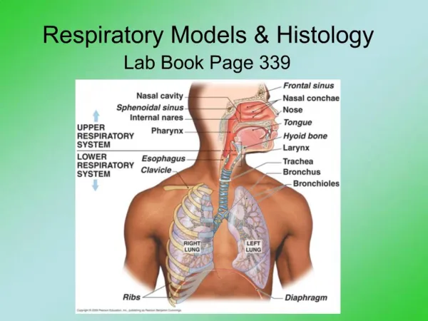
Histology of Respiratory System. Respiratory System. Conducting Part -responsible for passage of air and conditioning of the inspired air. Examples: nasal cavities,pharynx, trachea, bronchi and their intrapulmonary continuations.

E N D
Respiratory System • Conducting Part-responsible for passage of air and conditioning of the inspired air. Examples:nasal cavities,pharynx, trachea, bronchi and their intrapulmonary continuations. • RespiratoryPart-involved with the exchange of oxygen and carbondioxide between blood and inspires air.Includes the lungs
RESPIRATORY SYSTEM HISTOLOGY • Trachea • Bronchus -Primary bronchus -Secondary bronchus -Tertiary bronchus • Bronchiole • Lung
Trachea • Mucosa -Epithelium -Lamina propria • Sub mucosa • Cartilage &muscle layer • Adventitia
Trachea Mucosa • Epithelium -Pseudo stratified ciliated columnar/Respiratory epithelium Cells-Ciliated columnar cells - Goblet cells -Brush cells - Basal cells -Granule (kulchitsky) cells -Clara cells( bronchiolar cells) surfactant secretion • Lamina propria - Elastic fibre, Lymphocyte, Mast cells, Blood vessels
Trachea • Sub mucosa- • Loose connective tissue • Tracheal glands-Mixed (serous &mucus) glands • Blood vessels and ducts • Cartilage &smooth muscle layer- • ”C” Shaped hyaline cartilage having perichondrium and chondrocytes • Ends of cartilage connected by smooth muscles • Adventitia-fibro elastic tissue
Bronchus • Principal bronchus -same as trachea • Secondary /Lobar • bronchus -Irregular hyaline cartilage -Pseudo stratified ciliated columnar • Tertiary /Segmental bronchus -Columnar epithelium -Patches of cartilage
Changes as bronchi become smaller • Cartilage-irregular and smaller. Absent in bronchioles. • Muscle- increases as bronchi becomes smaller.(Spasm of these muscles bring difficulty in breathing in allergic conditions) • Subepithelial Lymphoid Tissue-increases with decrease in the diameter of bronchi. • Glands-few.Absent in the walls of capillaries. • Epithelium- pseudostratified ciliated columnar epithelium in principal bronchi later simple ciliated columnar,non-ciliated columnar and later cuboidal in respiratory bronchioles
Bronchiole • Terminal bronchiole -Columnar epithelium -No cartilage - smooth muscle + -Clara cells present • Respiratory bronchiole -Cuboidal epithelium -No mucous gland
Differences between Bronchi and Bronchioles Bronchioles • No glands • No cartilage • No goblet cells • Thick smooth muscle layer • Presence of Clara cells • Many elastic fibres
Cells seen in the respiratory passages • Goblet cells • Non-ciliated serous cells • Basal cells • Cells of Clara • Brush cells • Argyrophil Cells similar to diffuse endocrine cells of gut • Lymphocytes
Goblet cells: numerous and secrete mucous. Mucous traps the dust particles and is moved by ciliary action towards pharynx. • Non-ciliated serous cells: secretes watery fluid that keeps the epithelium moist • Cells of Clara: are non-ciliated cells predominantly seen in terminal bronchioles. Secrete a fluid that spreads over the alveolar surface forming a film that reduces surface tension. May function as stem cells
Basal cells: Multiply and transform into other cell types replace the lost cells. • Argyrophil cells: cells similar to diffuse endocrine cells of the gut containing granules, secrete hormones and active peptides including serotonin and bombesin. • Lymphocytes and other leucoctes may be present in the epithelium.
Alveoli • 200 million in a normal lung • Total area-75 square meters • Total capillary surface area available for exchange-125square meters • Are spongy and form the parenchyma of lung. • Sac like evaginations present at the terminal end of the bronchial tree.
In section, they resemble a honeycomb • Alveoli are separated by interalveolar septum lying between thin epithelial lining of two neighbouring alveoli • Interalveolar septum contains anetwork of capillaries supported by reticular and elastic fibres, occassionally fibroblasts, macrophages and mast cells. • Septum containspores(ALVEOLAR PORES OF KOHN) help in passage of air from one alveolus to another, thus equalizing Pressure in the alveoli
Elastic fibres-enable the alveoli to expand during inspiration and passively contract during expiration. • Reticular fibres support and prevent overdistention of the alveoli
Cells in the Alveoli • Type I Pneumocytes • Type II Pneumocytes • Macrophages or Dust cells
Pneumocytes • Type I Alveolar or Type I Pneumocytes orSquamous Epithelial cells- Form the lining of 90% of the alveolar surface, numerous,squamous,thinness reduced to 0.05 to0.2 micron m, edges of the 2 cells overlap and are uniting by tight junctions- preventing leakage of blood from capillaries to the alveolar lumen • Form Blood Air barrier
Type II Alveolar or Type II pneumocytes • Also known as Septal cells • Rounded or cuboidal secretory cells with microvilli • Secretory granules are made of several layers- Multilamellar bodies. • These lamillar bodies are cytoplasmic inclusions made up of phospholipid which combines with other chemicals to form surfactant & then ooze out of the cell by exocytosis. • Pulmonary Surfactant – is the fluid secreted that spreads over the alveolar surface • These cells can multiply to replace damaged cells. • Surfactant also has bactericidal properties
Pulmonary Surfactant • Surfactant contains phospholipids, proteins and glycosaminoglycans, reduces the surface tension and prevents collapse of the alveolus during expiration. • Is constantly renewed. • Removed from the surface by Type I pneumocytes and macrophages • The reduced surface tension in the alveoli decreases the force that is needed to inflate alveoli during inspiration. • Therefore surfactant stabilizesthe alveolar diameters, facilitates their expansion and prevents their collapse by minimizing the collapsing forces
Blood Air Barrier • Consist of a thin layer of surfactant • Cytoplasm of Type I Pneumocytes • Basement membrane of Pneumocytes • Intervening Connective Tissue • Basement membrane of capillary endothelial cell • Cytoplasm of capillary Endothelial cells • Endothelial cells of alveolar capillaries are extremely thin, have numerous projections increasing the surface area of the cell membrane exposed to blood for gaseous exchange. At places the 2 basement membranes are so fused reducing the thickness of Barrier.
Alveolar Macrophages or Dust cells • Derived from Monocytes and are part mononuclear phagocytic system. • Either seen in the septa or alveoli • Cytoplasm contains phagocytosed inhaled carbon and dust particles • Inhaled carbon and dust particles are passed on to them from pneumocyte I through pinocytic vesicles