Download
1 / 85
870 likes | 1.11k Views
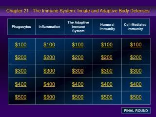
21. The Immune System: Innate and Adaptive:Body Defenses: Part A. Immunity. Resistance to disease Immune system has two intrinsic systems Innate (nonspecific) defense system Adaptive (specific) defense system. Immunity. Innate defense system has two lines of defense

E N D
21 The Immune System: Innate and Adaptive:Body Defenses: Part A
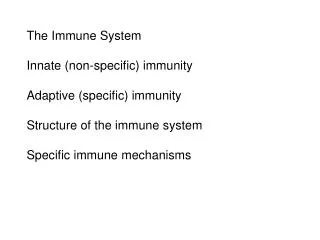
Immunity • Resistance to disease • Immune system has two intrinsic systems • Innate (nonspecific) defense system • Adaptive (specific) defense system
Immunity • Innate defense system has two lines of defense • First line of defense is external body membranes (skin and mucosae) • Second line of defense is antimicrobial proteins, phagocytes, and other cells • Inhibit spread of invaders • Inflammation is its most important mechanism
Immunity • Adaptive defense system • Third line of defense attacks particular foreign substances • Takes longer to react than the innate system • Innate and adaptive defenses are deeply intertwined
Surface barriers • Skin • Mucous membranes Innate defenses Internal defenses • Phagocytes • NK cells • Inflammation • Antimicrobial proteins • Fever Humoral immunity • B cells Adaptive defenses Cellular immunity • T cells Figure 21.1
Innate Defenses • Surface barriers • Skin, mucous membranes, and their secretions • Physical barrier to most microorganisms • Keratin is resistant to weak acids and bases, bacterial enzymes, and toxins • Mucosae provide similar mechanical barriers
Surface Barriers • Protective chemicals inhibit or destroy microorganisms • Skin acidity • Lipids in sebum and dermcidin in sweat • HCl and protein-digesting enzymes of stomach mucosae • Lysozyme of saliva and lacrimal fluid • Mucus
Surface Barriers • Respiratory system modifications • Mucus-coated hairs in the nose • Cilia of upper respiratory tract sweep dust- and bacteria-laden mucus from lower respiratory passages
Internal Defenses: Cells and Chemicals • Necessary if microorganisms invade deeper tissues • Phagocytes • Natural killer (NK) cells • Inflammatory response (macrophages, mast cells, WBCs, and inflammatory chemicals) • Antimicrobial proteins (interferons and complement proteins) • Fever
Phagocytes: Macrophages • Macrophages develop from monocytes to become the chief phagocytic cells • Free macrophages wander through tissue spaces • E.g., alveolar macrophages • Fixed macrophages are permanent residents of some organs • E.g., Kupffer cells (liver) and microglia (brain)
Phagocytes: Neutrophils • Neutrophils • Become phagocytic on encountering infectious material in tissues
Mechanism of Phagocytosis Step 1: Adherence of phagocyte to pathogen • Facilitated by opsonization—coating of pathogen by complement proteins or antibodies
Innate defenses Internal defenses (a) A macrophage (purple) uses its cytoplasmicextensions to pull spherical bacteria (green) toward it. Scanning electron micrograph (1750x). Figure 21.2a
1 Phagocyte adheres to pathogens or debris. 2 Phagocyte forms pseudopods that eventually engulf the particles forming a phagosome. Phagosome (phagocytic vesicle) Lysosome 3 Lysosome fuses with the phagocytic vesicle, forming a phagolysosome. Acid hydrolase enzymes 4 Lysosomal enzymes digest the particles, leaving a residual body. 5 Exocytosis of the vesicle removes indigestible and residual material. (b) Events of phagocytosis. Figure 21.2b
Mechanism of Phagocytosis • Destruction of pathogens • Acidification and digestion by lysosomal enzymes • Respiratory burst • Release of cell-killing free radicals • Activation of additional enzymes • Oxidizing chemicals (e.g. H2O2) • Defensins (in neutrophils)
Natural Killer (NK) Cells • Large granular lymphocytes • Target cells that lack “self” cell-surface receptors • Induce apoptosis in cancer cells and virus-infected cells • Secrete potent chemicals that enhance the inflammatory response
Inflammatory Response • Triggered whenever body tissues are injured or infected • Prevents the spread of damaging agents • Disposes of cell debris and pathogens • Sets the stage for repair
Inflammatory Response • Cardinal signs of acute inflammation: • Redness • Heat • Swelling • Pain (And sometimes 5. Impairment of function)
Inflammatory Response • Macrophages and epithelial cells of boundary tissues bear Toll-like receptors (TLRs) • TLRs recognize specific classes of infecting microbes • Activated TLRs trigger the release of cytokines that promote inflammation
Inflammatory Response • Inflammatory mediators • Histamine (from mast cells) • Blood proteins • Kinins, prostaglandins (PGs), leukotrienes, and complement • Released by injured tissue, phagocytes, lymphocytes, basophils, and mast cells
Vasodilation and Increased Vascular Permeability • Inflammatory chemicals cause • Dilation of arterioles, resulting in hyperemia • Increased permeability of local capillaries and edema (leakage of exudate) • Exudate contains proteins, clotting factors, and antibodies
Inflammatory Response: Edema • Functions of the surge of exudate • Moves foreign material into lymphatic vessels • Delivers clotting proteins to form a scaffold for repair and to isolate the area
Innate defenses Internal defenses Tissue injury Release of chemical mediators (histamine, complement, kinins, prostaglandins, etc.) Release of leukocytosis- inducing factor Leukocytosis (increased numbers of white blood cells in bloodstream) Vasodilation of arterioles Increased capillary permeability Attract neutrophils, monocytes, and lymphocytes to area (chemotaxis) Leukocytes migrate to injured area Local hyperemia (increased blood flow to area) Capillaries leak fluid (exudate formation) Margination (leukocytes cling to capillary walls) Initial stimulus Physiological response Signs of inflammation Diapedesis (leukocytes pass through capillary walls) Leaked clotting proteins form interstitial clots that wall off area to prevent injury to surrounding tissue Leaked protein-rich fluid in tissue spaces Result Phagocytosis of pathogens and dead tissue cells (by neutrophils, short-term; by macrophages, long-term) Heat Redness Pain Swelling Temporary fibrin patch forms scaffolding for repair Locally increased temperature increases metabolic rate of cells Possible temporary limitation of joint movement Pus may form Area cleared of debris Healing Figure 21.3
Phagocyte Mobilization • Neutrophils, then phagocytes flood to inflamed sites
Phagocyte Mobilization • Steps for phagocyte mobilization • Leukocytosis: release of neutrophils from bone marrow in response to leukocytosis-inducing factors from injured cells • Margination: neutrophils cling to the walls of capillaries in the inflamed area • Diapedesis of neutrophils • Chemotaxis: inflammatory chemicals (chemotactic agent) promote positive chemotaxis of neutrophils
Innatedefenses Internaldefenses Inflammatorychemicalsdiffusingfrom theinflamed siteact as chemotacticagents. 4 Chemotaxis.Neutrophilsfollow chemicaltrail. Capillary wall Basementmembrane Endothelium 1 2 3 Leukocytosis.Neutrophils enter bloodfrom bone marrow. Margination.Neutrophils clingto capillary wall. Diapedesis.Neutrophils flatten andsqueeze out of capillaries. Figure 21.4
Innatedefenses Internaldefenses Inflammatorychemicalsdiffusingfrom theinflamed siteact as chemotacticagents. Capillary wall Basementmembrane Endothelium 1 Leukocytosis.Neutrophils enter bloodfrom bone marrow. Figure 21.4, step 1
Innatedefenses Internaldefenses Inflammatorychemicalsdiffusingfrom theinflamed siteact as chemotacticagents. Capillary wall Basementmembrane Endothelium 1 2 Leukocytosis.Neutrophils enter bloodfrom bone marrow. Margination.Neutrophils clingto capillary wall. Figure 21.4, step 2
Innatedefenses Internaldefenses Inflammatorychemicalsdiffusingfrom theinflamed siteact as chemotacticagents. Capillary wall Basementmembrane Endothelium 1 2 3 Leukocytosis.Neutrophils enter bloodfrom bone marrow. Margination.Neutrophils clingto capillary wall. Diapedesis.Neutrophils flatten andsqueeze out of capillaries. Figure 21.4, step 3
Innatedefenses Internaldefenses Inflammatorychemicalsdiffusingfrom theinflamed siteact as chemotacticagents. 4 Chemotaxis.Neutrophilsfollow chemicaltrail. Capillary wall Basementmembrane Endothelium 1 2 3 Leukocytosis.Neutrophils enter bloodfrom bone marrow. Margination.Neutrophils clingto capillary wall. Diapedesis.Neutrophils flatten andsqueeze out of capillaries. Figure 21.4, step 4
Antimicrobial Proteins • Interferons (IFNs) and complement proteins • Attack microorganisms directly • Hinder microorganisms’ ability to reproduce
Interferons • Viral-infected cells are activated to secrete IFNs • IFNs enter neighboring cells • Neighboring cells produce antiviral proteins that block viral reproduction
Innate defenses Internal defenses Virus 1 Virusenters cell. New viruses Viral nucleic acid 5 Antiviralproteins blockviralreproduction. 2 Interferongenes switch on. DNA Nucleus mRNA 4 Interferonbindingstimulates cell toturn on genes forantiviral proteins. 3 Cell producesinterferonmolecules. Interferon Host cell 2Binds interferon from cell 1; interferon induces synthesis ofprotective proteins Host cell 1Infected by virus;makes interferon;is killed by virus Figure 21.5
Innate defenses Internal defenses Virus 1 Virusenters cell. Viral nucleic acid Nucleus Host cell 2Binds interferon from cell 1; interferon induces synthesis ofprotective proteins Host cell 1Infected by virus;makes interferon;is killed by virus Figure 21.5, step 1
Innate defenses Internal defenses Virus 1 Virusenters cell. Viral nucleic acid 2 Interferongenes switch on. DNA Nucleus Host cell 2Binds interferon from cell 1; interferon induces synthesis ofprotective proteins Host cell 1Infected by virus;makes interferon;is killed by virus Figure 21.5, step 2
Innate defenses Internal defenses Virus 1 Virusenters cell. Viral nucleic acid 2 Interferongenes switch on. DNA Nucleus mRNA 3 Cell producesinterferonmolecules. Interferon Host cell 2Binds interferon from cell 1; interferon induces synthesis ofprotective proteins Host cell 1Infected by virus;makes interferon;is killed by virus Figure 21.5, step 3
Innate defenses Internal defenses Virus 1 Virusenters cell. Viral nucleic acid 2 Interferongenes switch on. DNA Nucleus mRNA 4 Interferonbindingstimulates cell toturn on genes forantiviral proteins. 3 Cell producesinterferonmolecules. Interferon Host cell 2Binds interferon from cell 1; interferon induces synthesis ofprotective proteins Host cell 1Infected by virus;makes interferon;is killed by virus Figure 21.5, step 4
Innate defenses Internal defenses Virus 1 Virusenters cell. New viruses Viral nucleic acid 5 Antiviralproteins blockviralreproduction. 2 Interferongenes switch on. DNA Nucleus mRNA 4 Interferonbindingstimulates cell toturn on genes forantiviral proteins. 3 Cell producesinterferonmolecules. Interferon Host cell 2Binds interferon from cell 1; interferon induces synthesis ofprotective proteins Host cell 1Infected by virus;makes interferon;is killed by virus Figure 21.5, step 5
Interferons • Produced by a variety of body cells • Lymphocytes produce gamma (), or immune, interferon • Most other WBCs produce alpha () interferon • Fibroblasts produce beta () interferon • Interferons also activate macrophages and mobilize NKs
Interferons • Functions • Anti-viral • Reduce inflammation • Activate macrophages and mobilize NK cells • Genetically engineered IFNs for • Antiviral agents against hepatitis and genital warts virus • Multiple sclerosis treatment
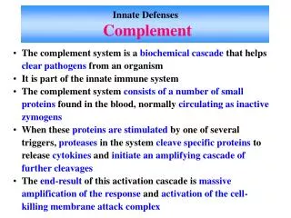
Complement • ~20 blood proteins that circulate in an inactive form • Include C1–C9, factors B, D, and P, and regulatory proteins • Major mechanism for destroying foreign substances
Complement • Amplifies all aspects of the inflammatory response • Kills bacteria and certain other cell types by cell lysis • Enhances both nonspecific and specific defenses
Complement Activation • Two pathways • Classical pathway • Antibodies bind to invading organisms • C1 binds to the antigen-antibody complexes (complement fixation) • Alternative pathway • Triggered when activated C3, B, D, and P interact on the surface of microorganisms
Complement Activation • Each pathway involves activation of proteins in an orderly sequence • Each step catalyzes the next • Both pathways converge on C3, which cleaves into C3a and C3b
Complement Activation • Activated complement • Enhances inflammation • Promotes phagocytosis • Causes cell lysis • C3b initiates formation of a membrane attack complex (MAC) • MAC causes cell lysis by inducing a massive influx of water • C3b also causes opsonization, and C3a causes inflammation
Alternative pathway Classical pathway Spontaneous activation Antigen-antibody complex + + Stabilizing factors (B, D, and P) + complex No inhibitors on pathogen surface Enhances inflammation: Opsonization: stimulates histamine release, increases blood vessel permeability, attracts phagocytes by chemotaxis, etc. coats pathogen surfaces, which enhances phagocytosis Insertion of MAC and cell lysis (holes in target cell’s membrane) Pore Complement proteins (C5b–C9) Membrane of target cell Figure 21.6
Fever • Systemic response to invading microorganisms • Leukocytes and macrophages exposed to foreign substances secrete pyrogens • Pyrogens reset the body’s thermostat upward
Fever • High fevers are dangerous because heat denatures enzymes • Benefits of moderate fever • Causes the liver and spleen to sequester iron and zinc (needed by microorganisms) • Increases metabolic rate, which speeds up repair
Adaptive Defenses • The adaptive immune (specific defense) system • Protects against infectious agents and abnormal body cells • Amplifies the inflammatory response • Activates complement
Adaptive Defenses • Adaptive immune response • Is specific • Is systemic • Has memory • Two separate overlapping arms • Humoral (antibody-mediated) immunity • Cellular (cell-mediated) immunity