Download
1 / 12
120 likes | 270 Views
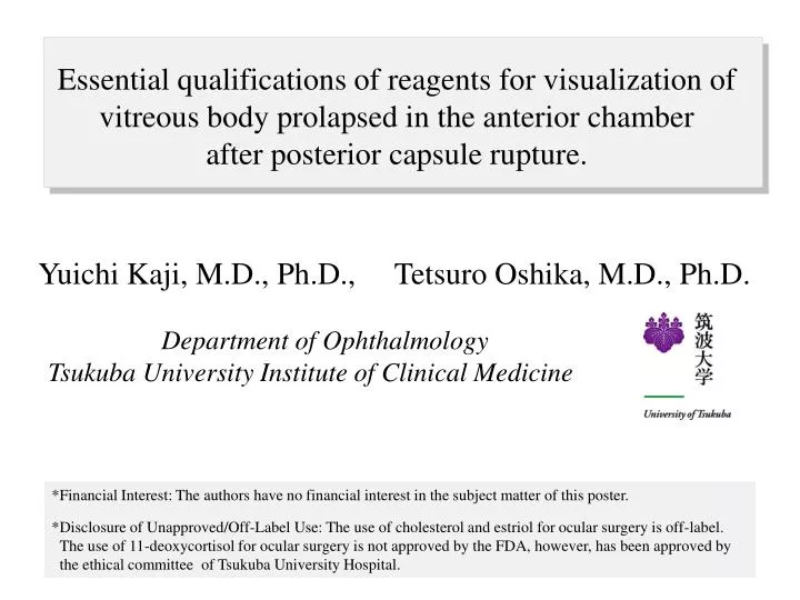
Essential qualifications of reagents for visualization of vitreous body prolapsed in the anterior chamber after posterior capsule rupture. Yuichi Kaji, M.D., Ph.D., Tetsuro Oshika, M.D., Ph.D. Department of Ophthalmology

E N D
Essential qualifications of reagents for visualization of vitreous body prolapsed in the anterior chamber after posterior capsule rupture. Yuichi Kaji, M.D., Ph.D.,Tetsuro Oshika, M.D., Ph.D. Department of Ophthalmology Tsukuba University Institute of Clinical Medicine *Financial Interest: The authors have no financial interest in the subject matter of this poster. *Disclosure of Unapproved/Off-Label Use: The use of cholesterol and estriol for ocular surgery is off-label. The use of 11-deoxycortisol for ocular surgery is not approved by the FDA, however, has been approved by the ethical committee of Tsukuba University Hospital.
Introduction Posterior capsule rupture with subsequent vitreous loss is one of the most common complications of cataract surgery.1,2 To minimize the post-operative complications after posterior capsule rupture, complete removal of prolapsed vitreous body in the anterior chamber is necessary.1,2 However, direct recognition of the vitreous body under surgical microscope is difficult, especially when the anterior chamber is filled with lens fragments and viscoelastic agents. To visualize the vitreous body during pars plana vitrectomy, an injection of a suspension of triamcinolone acetonide has been used.3 Triamicinolone acetonide can be used to visualize the vitreous body in the anterior chamber after posterior capsule rupture.4 However, triamcinolone acetonide left in the eye might be the cause of complications including ocular hypertension. In the present study, we compared various chemical reagents including triamcinolone acetonide for the safety and efficacy of visualization of vitreous body. 1. Chan FM, et al. Short-term outcomes in eyes with posterior capsule rupture during cataract surgery. J Cataract Refract Surg 29: 537-541, 2003. 2. Gimbel HV, et al. Intraoperative management of posterior capsule tears in phacoemulsification and intraocular lens implantation. Ophthalmology 108: 2186-2189, 2001. 3. Peyman GA, et al. Triamcinolone acetonide as an aid to visualization of the vitreous and the posterior hyaloid during pars plana vitrectomy. Retina20: 554-555, 2000. 4. Burk SE, et al. Visualizing vitreous using Kenalog suspension. J Cataract Refract Surg 29: 645-651, 2003.
Materials and Methods #0 #0-0: Preparation of the substances suspensions We compared the following 4 chemical reagents for the safety and efficacy of visualization of vitreous body in the anterior chamber using animal models. In addition, the suspension of the following reagents in balanced salt solution (BSS) were prepared. Triamcinolone Acetonide (Bristol-Myers, NY) Cholesterol (Sigma, St. Louis, MO) Estriol (Sigma, St. Louis, MO) 11-Deoxycortisol (Sigma, St. Louis, MO)
Materials and Methods #1 #1-1 Animal Model of Posterior Capsule Rupture Forty porcine eyes were purchased from a slaughterhouse. After making a corneoscleral wound and continuous curvilinear capsulorhexis, the lens was aspirated using a phaco machine. Then the posterior capsule was aspirated and torn off using an irrigation/aspiration tip, so that the vitreous body would prolapse into the anterior chamber. Suspensions of 10mg/ml of triamcinolone acetonide, cholesterol, estriol and 11-deoxycortisol in BSS were prepared. After intentionally rupturing the posterior capsule of the porcine eye, 0.5 ml of one of the suspensions was injected through the corneoscleral wound. An anterior vitrectomy system was utilized to remove the prolapsed vitreous body. After the gentle irrigation and aspiration, the non-adherent particles were removed from the anterior chamber. Then, the vitreous body entrapping the white granules of the respective substance was removed. Ten porcine eyes were used for each reagent.
Materials and Methods #2 Toxicity of the reagents injected in the anterior chamber. #2-1 Slit-Lamp Examination Suspensions of 5 mg/0.1ml of triamcinolone acetonide, cholesterol, estriol and 11-deoxycortisol in BSS were injected in the anterior chamber of New Zealand white raibbits. As controls, same volume of the BSS alone was injected into the left eye. No other surgical procedure was performed. #2-2 Intraocular Pressure In the eyes receiving injections in the anterior chamber, biomicroscopic examinations were given and intraocular pressure measurements taken using a pneumatic tonometer (MENTOR, model 30 classic, Norwell, MA) before and 12 hours, 1, 2, 3, 7, 14 and 28 days after the injection. #2-3 Corneal Endothelial Density The corneal endothelial cell density was measured using a contact specular microscope (KONAN medical, type class I, Hyogo, Japan) before and 28 days after the injection. #2-4 Histology Hematoxylin-Eosin staining of the corneas at 28 days after injection of the reagents into the anterior chamber was prepared.
Results #1 Immediate after Post-cap rupture After gentle irrigation/aspiration Injection Triamcinolone Acetonide Cholesterol Estriol 11-Deoxycortisol All the reagents were useful in visualization of vitreous body prolapsed in the anterior chamber after posterior capsule rupture.
Results #2-1 Immediate after Injection 12 hours 1 day 7 days Triamcinolone Acetonide Cholesterol Estriol 11-Deoxycortisol Cholesterol induced severe corneal edema and injection. In contrast, the other reagents disappeared from the anterior chamber within a day without any significant complications.
Results #2-2 Cholesterol Triamcinolone Acetonide Estriol 11-Dexycortisol Intraocular Pressure (mmHg) 0 1 2 3 7 14 28 days Cholesterol injected into the anterior chamber induced significant increase in the intraocular pressure. In contrast, the other reagents injected in the anterior chamber had no significant effect on the intraocular pressure.
Results #2-3 Corneal Endothelial Cell Density (/mm2) uncountable before before before before 28 days 28 days 28 days Triamcinolone Acetonide Cholesterol Estriol 11-Deoxycortisol Triamcinolone acetonide, estriol, and 11-deoxycortisol injected into the anterior chamber had no significant effect on the corneal endothelial cell density. In cotrast, corneal endothelial cell density could not evaluated because of severe corneal edema after injection of cholesterol.
Results #2-4 Triamcinolone Acetonide Cholesterol Estriol 11-Deoxycortisol Keratic precipitate containing macrophages, neutrophils and lymphocytes with neovascularization. Triamcinolone acetonide, estriol, and 11-deoxycortisol injected into the anterior chamber had no significant effect on the corneal structure. In cotrast, cholesterol induced loss of corneal endothelial cells, corneal stromal edema, severe infiltration of inflammatory cells in the anterior chamber.
Discussion Safety IOP Corneal Endothelium Histology Visualization of vitreous body. Triamcinolone Acetonide Estriol Cholesterol 11-Deoxycortisol Good Good Good Good no changes no changes Severe Toxicities no changes All the four reagents were effective in visualization of vitreous body in the anterior chamber after posterior capsule rupture. However, cholesterol injected into the anterior chamber had severe toxicities. In contrast, triamcinolone acetonide, estriol, and 11-deoxycortisol had no significant toxicities. For these reasons, estriol and 11-deoxycortisol in addition to triamicinolone acetonide could be used to visualize the vitreous body after posterior capsule rupture.
Summary hydrophilic modifications hydrophobic modifications Triamcinolone Acetonide (Bristol-Myers, NY) Cholesterol (Sigma, St. Louis, MO) Estriol (Sigma, St. Louis, MO) 11-Deoxycortisol (Sigma, St. Louis, MO) Cholesterol skeleton is necessary for an adherence to the vitreous body. All the four reagents have hydrophobic modifications. However, there is only one hydrophilic modifications in cholesterol. This is the reason why cholesterol injected into the anterior chamber tends to remain and induce ocular hypertension. In this way, molecules with cholesterol skeleton and several hydrophilic modifications are thought to be the essential qualifications of reagents for visualization of vitreous body without any complications. These are the molecular-based reasons why triamcinolone acetonide is effective in visualization of vitreous body in the anterior chamber after posterior capsule rupture.