Download
1 / 42
430 likes | 586 Views
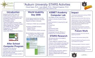
Auburn University MRI Research Center. Facility Overview. Three story – 45,000 SF building MRI systems 3 Tesla (T) open-bore whole body 7T whole body Designed to support research and clinical operations Animal facilities Short-term holding facility Minimally-invasive procedures area

E N D
Facility Overview • Three story – 45,000 SF building • MRI systems • 3 Tesla (T) open-bore whole body • 7T whole body • Designed to support research and clinical operations • Animal facilities • Short-term holding facility • Minimally-invasive procedures area • Training for faculty/staff/students • Funded by a $21M State bond issued in 2007
Auburn University Objectives • Increase AU research funding • Establish AU as a leader in MRI research • Enhance AU’s reputation as a major research university • Attract businesses/industries to the AU Research Park • Provide additional healthcare capabilities for the region
AU MRI Research Center • Partnership between • Auburn University • Siemens Healthcare • East Alabama Medical Center • Alabama Imaging Inc. • University of Alabama at Birmingham (UAB) • International • Central South University (China) • Siemens Corporate Technology (China)
Second Floor USAARL AU Research The Orthopaedic Clinic
Third Floor Rehab Works – EAMC AU Kinesiology Auburn Neurosurgery
Proposed Operating Scenario • EAMC/AI controls 3T and clinical area • 8 hours/day (8AM-4PM) Mon-Fri • Lease at market value • AU controls 3T • 16 hours/day (4PM-8AM) Mon-Fri • 24 hours/day Sat, Sun, holidays • 7T is research only • AU control 24/7
Timeline • June ‘09 – Order equipment • July ‘09 – Building design • July ‘09 – Begin seminar series • Jan ‘10 – Begin construction • Summer ‘10 – Begin training • Sept ‘10 – Construction complete • Oct ‘10 – 3T/7T install • Oct ‘10 – Begin 3T operations • Spring ‘11 – Begin 7T operations
Current and Potential Research Projects • Coil design & Manufacture • Technology commercialization • Wireless technology • Orthopedic Injury/Rehab • Auburn Athletics / Andrews Orthopedics • fMRI/Cognitive Neuroscience • Autism • Psychology / Human Sciences • Communication Disorders • Audiology • Speech Pathology • Advanced cardiac imaging • UAB collaboration (NIH) • Fat Imaging • Diabetes and obesity • Metabolic/Molecular imaging • MRS/MRSI • Alzheimer’s disease (NIH)
3T Open-Bore MRI • FDA certified for clinical use • 70 cm bore • 173 cm length
Standard Applications • Neurology • Angiography • Cardiac • Body • Oncology • Orthopedic • Pediactric • Scientific
Spine Imaging at 3T • 4-channel neck coil • 24-channel spine coil
Advanced Applications - DESS • Increased cartilage/fluid contrast
Advanced Neuro Applications • fMRI acquisition/analysis • Diffusion • Perfusion • BOLD Imaging • Advanced Functional Neuro
MAGNETOM UHF Community (22 sites): • Massachusetts General Hospital, Boston, USA • Imaging since 08/2002 • Institute for Neuro-Biology, Magdeburg, Germany • Imaging since 02/2005 • New York University, NYC, USA • Imaging since 03/2005 • Center for MR Research, Minneapolis, USA • Imaging with Siemens console since 12/2005 • Gachon Medical School, Seoul, Korea • Imaging since 04/2006 • Oregon Health, Portland, USA • First images 12/06 • Erwin Hahn Institute for Magnetic Resonance, Essen, Germany • Imaging since 09/2006 • Center for Imaging in Biomedicine, Lausanne, Switzerland • Imaging since 11/2006 • Max Plank Institute, Tübingen, Germany (9.4T) Imaging since 07/2007 • CEA, Paris, France Imaging since 02/2007 • CEA, Paris, France, 11.7T to be installed 2013 12. UPMC, Pittsburgh, USA • former GE system converted to Siemens • Imaging since 05/2007 13. Max Plank Institute, Leipzig, Germany • Imaging since 07/2007 14. Medical University of Vienna, Vienna, Austria • Imaging since 04/2008 15. DKFZ, Heidelberg, Germany • Imaging since 08/2008 16. Forschungszentrum Jülich, Germany (9.4T/900) • Imaging since 17. HUP, Philadelphia, PA, USA • Imaging since 06/2008 18. MDC, Berlin, Germany • Imaging since 12/2008 19. CAS, Beijing, China • To be installed in Fall 2009 20. Oxford University, Oxford, UK • To be installed in spring 2010 21. Auburn University, Auburn, Alabama, USA • To be installed Fall 2010 22. CMRR, Minneapolis, USA • To be installed in fall 2010
Other Siemens 7T Installations in USA • MGH/ Harvard/ MIT • NYU • Penn • Minnesota • Portland, Oregon • Pittsburgh
High-Field Imaging T2-weighted imaging of the human hippocampus 1.5T 7.0T
fMRI at 1.5T, 3.0T, 7.0T Van der Zwaag, et al, Neuroimage. 2009 May 14
fMRI at 1.5T, 3.0T, 7.0T Van der Zwaag, et al, Neuroimage. 2009 May 14
7.0T 1.5T Behr, et al, Skeletal Radiol. 2009 Sep;38(9):911-7
7.0T 1.5T Behr, et al, Skeletal Radiol. 2009 Sep;38(9):911-7
Spatial Resolution • Regatte, et al, JMRI Vol.25, 2 Pages: 262-269 • Kuhl, et al, Radiology. 2008 Apr;247(1):16-35 • Yacob, et al, Neuroimage. 2007 October 1; 37(4): 1161–1177 • Nakada, Brain and Development ,29 (6), July 2007, 325-335 • Thomas, et al, JMRI 2008 November; 28(5): 1266–1272 • Miller, et al, J Neurophysiol 99:1969-1982, 2008 • Wiggins, et al, Magn Reson Med, 56(1), 216-223, 2006
Spectroscopy • Proton spectroscopy (3T and 7T) • Single voxel • Chemical shift imaging • Multinuclear spectroscopy (7T only) • 7Li, 13C, 17O, 19F, 23Na, 31P