Download
1 / 7
70 likes | 221 Views
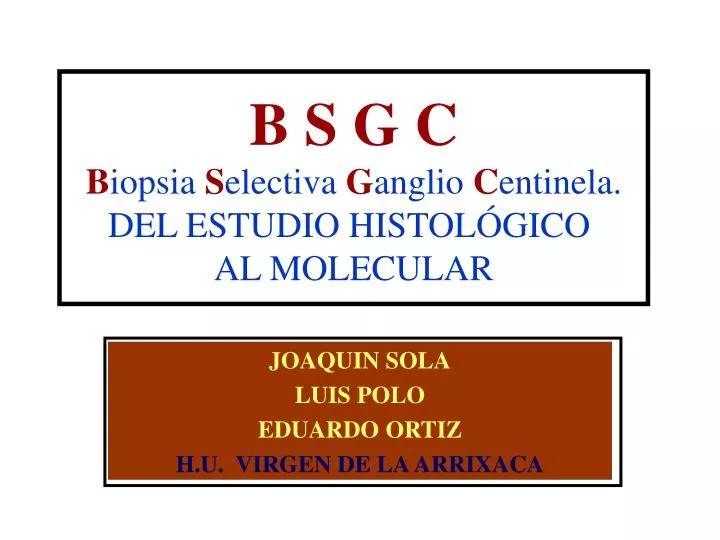
B S G C B iopsia S electiva G anglio C entinela. DEL ESTUDIO HISTOLÓGICO AL MOLECULAR. JOAQUIN SOLA LUIS POLO EDUARDO ORTIZ H.U. VIRGEN DE LA ARRIXACA. DEFINICION. Ganglio centinela es el primer ganglio o ganglios de una cadena linfática que recibe el

E N D
B S G CBiopsia Selectiva Ganglio Centinela. DEL ESTUDIO HISTOLÓGICO AL MOLECULAR JOAQUIN SOLA LUIS POLO EDUARDO ORTIZ H.U. VIRGEN DE LA ARRIXACA
DEFINICION Ganglio centinela es el primer ganglio o ganglios de una cadena linfática que recibe el flujo linfático desde el tumor primario.
SELECCION DE PACIENTES (ASCO 2005) • T1 – T2 ( hasta 3 cm.) • CDIS bg extenso (> 4cm.) • CDIS ag (> 2,5 cm.) • Carcinoma multicéntrico • Ancianos. • Obesos. • Varón. • Pre-neoadyuvancia (fase III)
BASES DEL METODO DE BSGC • El cáncer de mama se extiende a un/unos ganglio/s antes de afectar al resto de ganglios axilares. • Si el GC esta libre de metástasis la probabilidad de afectación de los GNC es < 0,1%. • La aparición de complicaciones o morbilidad es mucho menor. • El examen histológico es el mejor método para detectar metástasis GC.
CLASIFICACION MORFOLOGICA DE LAS CELULAS TUMORALES GC. • Metástasis. (>2 mm) • mM: microMetástasis. (>0,2 mm - <2 mm) • CTAs: Células Tumorales Aisladas (< 0,2mm) • CTDi: Células Tumorales Dispersas (> 2mm) • CTDe: Células Tumorales Desplazadas (< 0,2 mm)
CLASIFICACION MORFOLOGICA DE LAS CEL. TUMORALES EN GANGLIO CENTINELA M Mm CTAs CTDi CTDe