Download
1 / 2
20 likes | 78 Views
A retina specialist will examine your eye to detect a macular hole. This is a specialized scan of the back of the eye using Optical Coherence tomography (OCT).

E N D
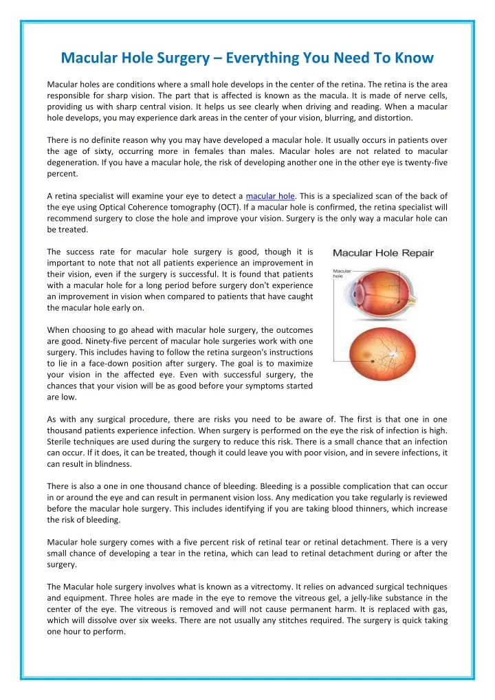
Macular Hole Surgery – Everything You Need To Know Macular holes are conditions where a small hole develops in the center of the retina. The retina is the area responsible for sharp vision. The part that is affected is known as the macula. It is made of nerve cells, providing us with sharp central vision. It helps us see clearly when driving and reading. When a macular hole develops, you may experience dark areas in the center of your vision, blurring, and distortion. There is no definite reason why you may have developed a macular hole. It usually occurs in patients over the age of sixty, occurring more in females than males. Macular holes are not related to macular degeneration. If you have a macular hole, the risk of developing another one in the other eye is twenty-five percent. A retina specialist will examine your eye to detect a macular hole. This is a specialized scan of the back of the eye using Optical Coherence tomography (OCT). If a macular hole is confirmed, the retina specialist will recommend surgery to close the hole and improve your vision. Surgery is the only way a macular hole can be treated. The success rate for macular hole surgery is good, though it is important to note that not all patients experience an improvement in their vision, even if the surgery is successful. It is found that patients with a macular hole for a long period before surgery don't experience an improvement in vision when compared to patients that have caught the macular hole early on. When choosing to go ahead with macular hole surgery, the outcomes are good. Ninety-five percent of macular hole surgeries work with one surgery. This includes having to follow the retina surgeon's instructions to lie in a face-down position after surgery. The goal is to maximize your vision in the affected eye. Even with successful surgery, the chances that your vision will be as good before your symptoms started are low. As with any surgical procedure, there are risks you need to be aware of. The first is that one in one thousand patients experience infection. When surgery is performed on the eye the risk of infection is high. Sterile techniques are used during the surgery to reduce this risk. There is a small chance that an infection can occur. If it does, it can be treated, though it could leave you with poor vision, and in severe infections, it can result in blindness. There is also a one in one thousand chance of bleeding. Bleeding is a possible complication that can occur in or around the eye and can result in permanent vision loss. Any medication you take regularly is reviewed before the macular hole surgery. This includes identifying if you are taking blood thinners, which increase the risk of bleeding. Macular hole surgery comes with a five percent risk of retinal tear or retinal detachment. There is a very small chance of developing a tear in the retina, which can lead to retinal detachment during or after the surgery. The Macular hole surgery involves what is known as a vitrectomy. It relies on advanced surgical techniques and equipment. Three holes are made in the eye to remove the vitreous gel, a jelly-like substance in the center of the eye. The vitreous is removed and will not cause permanent harm. It is replaced with gas, which will dissolve over six weeks. There are not usually any stitches required. The surgery is quick taking one hour to perform.
About Us: Mahi Muqit ophthalmologist, cataract, and vitreoretinal surgeon at two private clinics in London, United Kingdom. He provides patients with superior service and support with a range of surgical procedures to meet their eyesight requirements. He has built up a solid reputation for his eye services in the London area as an expert eye doctor and surgeon offering surgical retina, medical retina, and complex cataract surgery. He also offers surgery to patients suffering from diabetic retinopathy. Mahi Muqit is a member of the Royal College of Ophthalmologists, a member of the British and Eire Association of Vitreoretinal Surgeons, and the UK and Ireland Society of Cataract and Refractive Surgeons. To find out more, visit https://www.retinasurgeon.uk.com/. is a leading consultant
