Download
1 / 51
530 likes | 782 Views
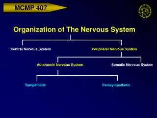
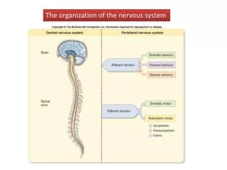
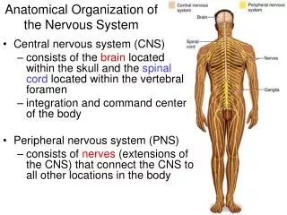
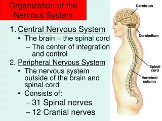
General Organization of the Nervous System. Parts. Central Nervous System : Brain and spinal cord Peripheral Nervous System : peripheral nerves, autonomic ganglia and dorsal root ganglia of the spinal cord. The Peripheral Nervous System. Somatovisceral sensory pathways

E N D
Parts • Central Nervous System: Brain and spinal cord • Peripheral Nervous System: peripheral nerves, autonomic ganglia and dorsal root ganglia of the spinal cord.
The Peripheral Nervous System • Somatovisceral sensory pathways • Somatic motor pathways to skeletal muscle • Autonomic motor pathways to visceral organs, skin and circulatory system. We are going to use the somatovisceral pathway as our initial way of looking at the peripheral NS
In order to process sensory information, the CNS must have answers to 3 questions: • What? Sensory receptors respond to specific stimulus modalities (touch, pressure, vibration, pain, temperature, light, chemicals, etc. • Where? CNS is organized as a set of maps showing the location of receptors – somatotopic organization. Each receptor, and each higher order neuron, has a discrete receptive field on the body surface, etc. • How much? In the neural code, stimulus intensity is encoded in action potential frequency
Basic Principles of Sensory Physiology • 1. a threshold level of intensity is required to initiate a sensory signal • Steps: • Stimulus causes an intensity-graded receptor (generator) potential within the sensory cell’s input zone • Receptor potential spreads to initial segment of axon by decremental conduction • Depending on the intensity of the stimulus, RP initiates no AP, 1 AP, or a burst or train of APs at the initial segment
2. Many receptors show adaptation: a decrease in responsiveness with continued stimulation Adaptation biases the receptor, making it more responsive to change in intensity versus steady unchanging stimuli. 3. The relationship between stimulus intensity and action potential frequency may be linear or logarithmic or some mixture of the two. Logarithmic responses are typical of receptors that address large variations in stimulus intensity
The somatovisceral sensory system • Processes information from the body surface, musculoskeletal system and body interior.
Merkel touch domes Ruffini’s corpuscles Meissner’s corpuscles Hair follicle receptors Pacinian corpuscles Steady pressure Steady pressure to low freq. vibration 30-40 Hz vibration Hair bending High frequency vibration (50-500 Hz) 5 types of Cutaneous Receptors
Receptive Field = area on the body surface within which an appropriate stimulus evokes a response. Notice the large difference in the sizes of the receptive fields of the 4 types of cutaneous receptors shown here. Arrows indicate direction-specificity
Other somatovisceral receptors • Thermoreceptors (2-3 types with different temp ranges) • Chemoreceptors (O2, CO2, osmolarity, etc) • Nociceptors (pain, discomfort) • Baroreceptors (blood pressure) • Muscle spindles (contain rapidly adapting and slowly adapting stretch receptors) • Golgi tendon organs
Organization of the Somatic Sensory System • Embryonic body segments (somites) are each served by a pair of spinal nerves from the corresponding spinal segment, or by a pair of cranial nerves from the corresponding head somite. • Each somite generates a dermatome that gives rise to skin covering and a myotome that gives rise to muscle. • Receptors that arise in the somite project to the corresponding spinal segment and its nearest neighbors.
The outcome of the body’s segmental organization is that each of the 30 pairs of spinal nerve roots serves an adjacent stripe of skin surface and a roughly corresponding block of skeletal muscle. • A similar process operates for the 3 branches of the trigeminal nerve versus the face.
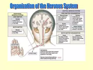
The somatic sensory pathways to the spinal cord Spinal nerves are mixed nerves containing both afferent and efferent axons. The afferents and efferents get separated near the cord so that the afferents pass through the dorsal root, where their cell bodies are, and the efferents pass out from the cord through the ventral horn. In the spinal cord, axons (white matter) are superficial to the cell bodies (gray matter).
Spinal afferents are divisible into 4 groups based on size and myelination • Group I: 13-20 mu diameter, myelinated, conduct. velocity 70-110 m/sec, carry muscle length information • Group II 6-12 mu diameter, myelinated, 25-70 m/sec conduction velocity, carry info from muscle, tendon receptors; Ruffini and Pacinian corpuscles • Group III 1-5 mu diameter, myelinated, 3.5-20 m/sec conduction velocity, slowly adapting touch/pressure receptors; “fast” nociceptors. • Group IV 1mu or less in in diameter, not myelinated, <1 m/sec conduction; “slow” pain, temperature, itch, tickle, etc.
Some authorities use a different form of categorization based on conduction velocity • Aβ: large, myelinated: mechanical stimuli • Aδ: small, myelinated: cold, fast pain, mechanical stimuli • C: small, unmyelinated: slow pain, temperature, mechanical stimuli
Spinal nerves are mixed nerves that contain both afferent and efferent axons. At the spinal cord most afferent axons split off dorsally and pass through the dorsal root ganglion where they connect with their cell bodies. Motor axons turn ventrally and enter the spinal cord through the ventral horn of the spinal segment. Note that unlike the brain, where the cell bodies are on the surface, the spinal gray matter is in the center and the white matter consisting of axons is on the outside.
Generally, each half-brain controls the contralateral body half • Spinal pathways decussate – a decussation occurs when a fiber tract crosses the midline while ascending or descending • A commissure is a connection between homologous structures on the two sides of the brain – so the corpus callosum is a bundle of fibers that represents the main route of communication between the two halves of the cortex. If this connection is severed, the patient can be shown to possess two minds that can communicate only by watching each other’s actions.
Two main spinal pathways carry somatovisceral sensations to the brain • 1. The dorsal column pathway: ascends on ipsilateral side of the spinal cord; decussates in the medulla of the brainstem; passes through thalamus to the contralateral somatosensory cortex. Good somatotopic organization is preserved throughout this pathway.
Note that the afferents in the dorsal column pathway don’t synapse on interneurons until they reach the dorsal column nuclei of the medulla. These interneurons then decussate to the contralateral medulla and ascend to the thalamus, where they synapse on 3rd order cells. Sensory modalities carried in dorsal column: cutaneous vibration, pressure, joint position, muscle stretch; visceral distention.
Note that higher order cells in the dorsal column system typicallyhave annular or bar-shaped receptive fields characterized by central excitatory zones surrounded by inhibitory zones. This arrangement is called lateral inhibition. It sharpens the system’s ability to detect stimulus location.
In lateral inhibition, more intense stimuli can cancel out the effects of less intense stimuli that arrive nearby. This biases the map of stimulus location so that edges of the stimulus show up much more than areas of no intensity contrast.
The pathways that mediate pain and temperature sense enter anterolateral fiber tracts. These pathways: Synapse on 2nd order neurons in the ipsilateral substantia gelatinosa of the spinal gray. The 2nd order neurons decussate in the same or adjacent segments of the spinal cord and thus ascend to the brain on the contralateral side of the cord.
As pain information ascends through the medulla, it provides important excitatory input to the brain’s reticular activating system, which has widespread connections throughout the brain and controls alertness and awareness.
Positional information is more highly conserved in the dorsal column system than in the anterolateral system As axons enter the dorsal columns, the ones from each spinal segment tend to stay together, so that positional information is preserved and the somatosensory cortex has an accurate map of the input from those receptors. In contrast, the axons that enter anterolateral pathways do not preserve positional information nearly as well, so sensations of pain and irritation tend to be more diffuse and poorly localized.
Information carried in ventrolateral tracts is filtered as it ascends the spinal cord. • Centrifugal (descending) pathways synapse on ascending neurons, releasing neuromodulators that inhibit release of excitatory transmitters in the ascending pathways – frequently these are endogenous opioids, accounting for the analgesic effect of opioid drugs and for stress-induced analgesia.
Spinal anatomy has diagnostic significance: Unilateral lesions of the spinal cord will interrupt specific types of information from each side of the body inferior to the lesion. Identification of the specific sensory deficits can be used to localize the spinal lesion.
Functional specialization of brain regions • Cerebrum • Cerebral cortex – perception, control of voluntary movement, consciousness • Basal nuclei – control of skeletal muscles • Limbic system – emotion and memory consolidation • Diencephalon – • Thalamus – relay station for sensory and motor information • Hypothalamus – homeostasis and behavioral drives • Cerebellum – coordinated body movements • Brainstem • Midbrain – ocular reflexes • Pons-medulla – involuntary functions including breathing, cardiovascular control, control of thoracic-abdominal organs
The cortex is divided into 4 lobes In some – but not all mammals, the cortex is thrown into folds called gyri (gyrus) separated by crevices called sulci (sulcus).
Sagittal section reveals some internal structures of the brain
Different brain regions have different evolutionary histories: The brainstem and cerebellum of mammals correspond fairly closely to those of reptiles. The limbic system – sometimes called the rhinencephalon or smelling brain – attained its present state relatively early in evolution of the mammalian line. Cortical structures are present to some extent in all mammals, but the volume and complexity of the cortex reaches a maximum in primates, in which the frontal parts of the cortex are referred to as neocortex because of their evolutionary newness – the olfactory cortex is older and has closer affinities with the limbic system.
The cortex typically has a columnar organization • For example, each of the whiskers (vibrissae) on the muzzle of a rat maps to a corresponding vertical cortical column on the face part of the map of the contralateral sensory cortex. Within this column are neurons that are activated specifically by vibrations of different frequencies, or by directional bending of the vibrissa.
This figure shows how receptors in the face of a rat map to neurons in the brainstem, the thalamus, and the somatosensory cortex. Because the positional arrangement of the afferent neurons closely follows the arrangement of receptive fields, the cortex has a very good representation of the face. This is another example of somatotopic organization.
Cortical organization is plastic (sometimes) • Plasticity refers to the power of the environment to shape the brain’s organization • Surgical ablation of individual vibrissae in a fetal animal results in the loss of the corresponding cortical column • Even in adulthood, loss of an appendage results in contraction of the corresponding part of the sensory cortical map, and increased use of a particular appendage results in an enlargement of its representation in the sensory cortex.
The cortex has a laminar organization • The cortex, an evolutionarily new part of the brain, is like a 6-story building with specific neuron types located on each floor. In the sensory cortex, sensory information arrives from the thalamus at the ground floor, where it is sorted out into submodalities. It then passes upward in columns that are each devoted to a single submodality, and is analyzed at increasingly greater detail as it passes to higher floors. Within this column are pyramidal cells, the main carriers of excitatory output from the cortex to other parts of the CNS. Synaptic inputs from interneurons in the cortical layers determine the ultimate output carried by the pyramidal cells.
Cortical neurons have complex input segments On the left, some typical pyramidal cells from different cortical regions. The purple bodies are the nuclei of other cells that didn’t get stained with the silver Golgi stain: some are neurons, some are glia.
The primary somatosensory cortex occupies a band posterior to the Fissure of Rolando and superior to the Sylvian Gyrus. Keep in mind that, because all somatosensory pathways arising below the neck decussate at some point, the left somatosensory cortex receives input from the contralateral side of the body and vice-versa.
The somatosensory cortex is organized as a map of the body surface The map, called the somatosensory homunculus, is distorted because areas of the body surface characterized by high receptor density are served by more cortical neurons See the next slide for a more up-to-date version
It turns out that common textbook versions of the somatosensory homunculus arecharacterized by some amount of prudery
Each sensory modality has primary and secondary (association) cortical regions
The primary motor cortex also has a somatotopic organization Notice the large amount of motor cortex that is devoted to the hand and face – these are the parts of the body over which we have the most sophisticated control. The situation would be different in quadrupeds or in animals in which facial expressions are less important.
Wernicke’s A. and Broca’s A. are the language areas of the cortex Damage to either one of these areas or to their connections leads to some form of aphasia. In the case of damage to Wernicke’s Area, the result is fluent but largely meaningless language. Such patients also have difficulty interpreting language. Damage to Broca’s area leads to halting, but logically intact speech. Language interpretation is not affected.
Interior Structures of the Brain • Limbic System • Thalamus • Basal Nuclei – we will learn about these in the context of somatic motor control • Hypothalamus
The limbic system consists of all of those structures shown in blue. In particular, the hippocampus is involved in formation and recovery of memory traces. In general, the limbic system plays a major role in emotional states such as anger and fear.
The hypothalamus is responsible for many homeostatic functions • Thermoregulation • Cardiovascular regulation • respiration • Body fluid volume/composition • Growth • Sexual differentiation and function • Motivation (“pleasure centers”) • Rage • Food intake and nutrient balance • Circadian rhythm
The thalamus is a relay station for sensory pathways leading to the cortex This is a repeat of an earlier slide
Cranial Nerves • connect to specialized sensory structures (eye, nose, ear, gustatory receptors): I= olfactory, II=optic, VIII=vestibuloauditory, gustatory = VII, IX, X) • Carry parasympathetic efferents to thorax/abdomen and visceral afferents to brainstem (X = vagus)