Download
1 / 58
580 likes | 587 Views
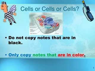
Explore the various components of cells, including the extracellular materials, cytoskeleton, membrane permeability, active transport, vesicular transport, membrane potential, cell adhesion molecules, membrane receptors, and the nucleus.

E N D
Extracellular Materials • Body fluids and cellular secretions • Extracellular matrix
Cytoskeleton • The “skeleton” on the cell • Dynamic, elaborate series of rods running through the cytosol • Consists of microtubules, microfilaments, and intermediate filaments
Cytoskeleton Figure 3.22
Effect of Membrane Permeability on Diffusion and Osmosis Figure 3.7a
Effect of Membrane Permeability on Diffusion and Osmosis Figure 3.7b
Passive Membrane Transport: Filtration • The passage of water and solutes through a membrane by hydrostatic pressure • Pressure gradient pushes solute-containing fluid from a higher-pressure area to a lower-pressure area
Tonicity – The ability os a solution to change the shape or tone of cells by altering its internal water volume • Isotonic – solutions with the same solute concentration as that of the cytosol • Hypertonic – solutions having greater solute concentration than that of the cytosol • Hypotonic – solutions having lesser solute concentration than that of the cytosol
Sodium-Potassium Pump Figure 3.9
Active Transport • Uses ATP to move solutes across a membrane • Requires carrier proteins
Types of Active Transport • Symport system – two substances are moved across a membrane in the same direction • Antiport system – two substances are moved across a membrane in opposite directions • Primary active transport – hydrolysis of ATP phosphorylates the transport protein causing conformational change
Types of Active Transport • Secondary active transport – use of an exchange pump (such as the Na+-K+ pump) indirectly to drive the transport of other solutes Figure 3.10
Types of Active Transport – Vesicular Transport • Transport of large particles and macromolecules across plasma membranes • Exocytosis – moves substance from the cell interior to the extracellular space • Endocytosis – enables large particles and macromolecules to enter the cell • Phagocytosis – pseudopods engulf solids and bring them into the cell’s interior
Vesicular Transport • Bulk-phase endocytosis – the plasma membrane infolds, bringing extracellular fluid and solutes into the interior of the cell • Receptor-mediated transport – uses clathrin-coated pits as the major mechanism for specific uptake of macromolecules
Vesicular Transport Pinocytosis Figure 3.12
Membrane Potential • Voltage across a membrane • Resting membrane potential • Ranges from –20 to –200 mV • Results from Na+ and K+ concentration gradients across the membrane • Differential permeability of the plasma membrane to Na+ and K+ Figure 3.13
Cell Adhesion Molecules (CAMs) • Anchor cells to the extracellular matrix • Assist in movement of cells past one another • Rally protective white blood cells to injured or infected areas
Roles of Membrane Receptors • Contact signaling – important in normal development and immunity • Electrical signaling – voltage-regulated “ion gates” in nerve and muscle tissue • Chemical signaling – neurotransmitters bind to chemically gated channel-linked receptors in nerve and muscle tissue • G protein-linked receptors – ligands bind to a receptor which activates a G protein, causing the release of a second messenger, such as cyclic AMP
Operation of a G protein • An extracellular ligand (first messenger), binds to a specific plasma membrane protein • The receptor activates a G protein that relays the message to an effector protein Figure 3.14
Operation of a G protein • The effector is an enzyme that produces a second messenger inside the cell • The second messenger activates a kinase • The activated kinase can trigger a variety of cellular responses Figure 3.14
Cytoplasm • Cytoplasm – material between plasma membrane and the nucleus • Cytosol – largely water with dissolved protein, salts, sugars, and other solutes • Cytoplasmic organelles – metabolic machinery of the cell • Inclusions – chemical substances such as glycosomes, glycogen granules, and pigment
Cytoplasmic Organelles – Learn Page 85 • Specialized cellular compartments • Membranous • Mitochondria, peroxisomes, lysosomes, endoplasmic reticulum, and Golgi apparatus • Nonmembranous • Cytoskeleton, centrioles, and ribosomes
Nucleus • Nuclear envelope, nucleoli, and chromatin • Gene-containing control center of the cell • Contains the genetic library with blueprints for nearly all cellular proteins • Dictates the kinds and amounts of proteins to be synthesized
Nucleus Figure 3.26a
Chromatin • Threadlike strands of DNA and histones • Arranged in fundamental units called nucleosomes • Form condensed, barlike bodies of chromosomes when the nucleus starts to divide Figure 3.27
DNA Replication • The DNA unwinds from the nucleosome • Helicase untwists the DNA double helix into two complementary nucleotide chains (replication bubble) • Freed nucleotide strands serve as templates for semiconservative replication Figure 3.29
DNA Replication • DNA polymerase creates leading and lagging strands • Short lagging strands of DNA are spliced together by DNA ligase • Histones associate with DNA and form chromatids that are united by a centromere Figure 3.29
Information Transfer from DNA to RNA • DNA triplets are transcribed into mRNA codons by RNA polymerase • Codons base pair with tRNA anticodons at the ribosomes • Amino acids are peptide bonded at the ribosomes to form polypeptide chains • Start and stop codons are used in initiating and ending translation
Information Transfer from DNA to RNA Figure 3.35
Protein Synthesis • DNA serves as master blueprint for protein synthesis • Genes are segments of DNA carrying instructions for a polypeptide chain • Triplets of nucleotide bases form the genetic library • Each triplet specifies coding for an amino acid
Nucleoli • Dark-staining spherical bodies within the nucleus • Not membrane bound • Site of ribosomal subunit production • Ribosomes are the site where proteins are manufactured
Rough ER – previous slide • External surface studded with ribosomes • Manufactures all secreted proteins • Responsible for the synthesis of integral membrane proteins and phospholipids for cell membranes
Signal Mechanism of Protein Synthesis • mRNA – ribosome complex is directed to rough ER by a signal-recognition particle (SRP) • SRP is released and polypeptide grows into cisternae • The protein is released into the cisternae and sugar groups are added Figure 3.17
Transcription • Transfer of information from the sense strand of DNA to mRNA • mRNA is synthesized from DNA using transcription factors and RNA polymerase • Each DNA triplet codes for a corresponding 3-base sequence of RNA, called a codon • There are 64 different codons • Introns are removed from pre-mRNA to produce functional mRNA
Translation • Translation of DNA is coded through mRNA to an amino acid sequence (polypeptide) • Involves all three types of RNA – mRNA, tRNA, and rRNA • Occurs in the cytoplasm at the ribosomes
Translation Figure 3.32
Translation Figure 3.32 continued
Genetic Code Figure 3.33
Roles of the Three Types of RNA • Messenger RNA (mRNA) carries the genetic information from DNA in the nucleus to the ribosomes in the cytoplasm • Transfer RNAs (tRNAs) bound to amino acids base pair with the codons of mRNA at the ribosome to begin the process of protein synthesis • Ribosomal RNA (rRNA) is a structural component of ribosomes
Protein Degradation • Nonfunctional organelle proteins are degraded by lysosomes • Ubiquitin attaches to soluble proteins and they are degraded in proteasomes Figure 3.36
Cell Cycle • Interphase • Growth (G1), synthesis (S), growth (G2) • Mitotic phase • Mitosis and cytokinesis Figure 3.28
Interphase • G1 (gap 1) – metabolic activity and vigorous growth • G0 – cells that permanently cease dividing • S (synthetic) – DNA replication • G2 (gap 2) – preparation for division
Cell Division • Essential for body growth and tissue repair • Mitosis – nuclear division • Cytokinesis – division of the cytoplasm
Mitosis • Prophase • Metaphase • Anaphase • Telophase
Cytokinesis • Cleavage furrow formed in late anaphase by contractile ring • Cytoplasm is pinched into two parts after mitosis ends
Early and Late Prophase • Asters are seen as chromatin condenses into chromosomes • Nucleoli disappear • Centriole pairs separate and the mitotic spindle is formed
Early and Late Prophase Figure 3.30.1
Early and Late Prophase Figure 3.30.2
Metaphase • Chromosomes cluster at the middle of the cell with their centromeres aligned at the exact center, or equator, of the cell • This arrangement of chromosomes along a plane midway between the poles is called the metaphase plate
Metaphase Figure 3.30.3