Download
1 / 5
50 likes | 52 Views
Immunoprecipitation Workflow and Troubleshooting Tips

E N D
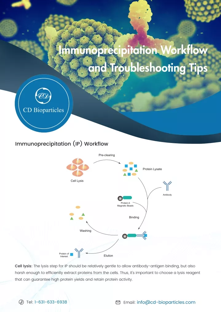
Immunoprecipitation Workflow and Troubleshooting Tips CD Immunoprecipitation (IP) Workflow Pre-clearing Protein Lysate Cell Lysis Antibody Protein A Magnetic Beads Binding Washing Protein of Interest Elution Cell lysis: The lysis step for IP should be relatively gentle to allow antibody-antigen binding, but also harsh enough to efficiently extract proteins from the cells. Thus, it's important to choose a lysis reagent that can guarantee high protein yields and retain protein activity. Email: info@cd-bioparticles.com Tel: 1-631-633-6938
Pre-clearing: This step reduces the number of non-specific contaminants in the cell lysate as well as removes proteins that possess a higher affinity to protein G or protein A before the specific IP steps. As a result of pre-cleaning, there would be less background noise and an improved signal-to-noise ratio. Binding: Magnetic beads (or agarose), antibody, and antigen form complexes at this step. The buffers used here and at the washing steps are vital to perform a successful IP. The order that these three components are added also affect the results. Antibodies may be added first to covalently or noncovalently bind to the magnetic beads before adding lysate, or incubated with lysate to form a complex before adding protein A or protein G magnetic beads to purify the complex from the mixture. Washing: Ideally, this step can break all nonspecific bindings without affecting the specific interactions between antibody and antigen. Adding extra lysis buffer to the washing buffer can help breaking the nonspecific bindings. Elution: Generally, there are three kinds of elution buffers, including glycine, SDS, and urea elution. Any of the three elution buffers can be used to elute the protein from the beads. The SDS buffer is harsh enough to elute noncovalently bound antibodies, antibody fragments, and the protein of interest. The glycine buffer is a gentler choice. Troubleshooting Tips 1. To improve elution conditions • Choose an appropriate lysis buffer according to the protein location (membrane, cytosolic, or nucleus). The pH, detergent, salt, and divalent cation concentrations should be optimized for each IP. • Check the antibody binding properties of each beads. It’s important to match the binding specificity of the beads to the species and the antibody subtypes (Table 1). Email: info@cd-bioparticles.com Tel: 1-631-633-6938
Table 1. Immunoglobulin binding properties. Species Immunoglobulin Isotype Protein A Protein G Protein A/G Human IgG1 +++ +++ +++ Human IgG2 +++ +++ +++ Human IgG3 + +++ +++ Human IgG4 +++ +++ +++ Human IgM + - + Human IgE ++ - ++ Human IgD - - - Human IgA + - + Human IgA1 + - + Human IgA2 + - + Human Fab + + + Human ScFv + - + Mouse IgG1 + ++ ++ Mouse IgG2a +++ +++ +++ Mouse IgG2b +++ +++ +++ Mouse IgG3 +++ +++ +++ Mouse IgM - - - Rat IgG1 + ++ ++ Rat IgG2a - +++ +++ Rat IgG2b - + + Rat IgG2c +++ +++ +++ Cow IgG1 + +++ +++ Cow IgG2 +++ +++ +++ Sheep IgG1 + +++ +++ Sheep IgG2 +++ +++ +++ Goat IgG1 + +++ +++ Goat IgG2 +++ +++ +++ Chicken IgY - - - Hamster IgG ++ ++ ++ Pig IgG +++ + +++ Horse IgG + +++ +++ Rabbit IgG +++ +++ +++ Cat IgG +++ + +++ Rhesus Monkey IgG +++ +++ +++ +++: Strong binding; ++: Medium binding; +: Weak binding; -: No binding. Email: info@cd-bioparticles.com Tel: 1-631-633-6938
• Alter the components, salt concentration, or pH of the elution buffer if no protein of interest is eluted from the beads. • Conduct a titration experiment first to optimize the antibody concentration if there isn’t enough antibodies for proper binding. • To enhance the expression of protein of interest, increase the volume of the cell lysate and pre-clearing the sample to decrease non-specific binding and remove debris. • Polyclonal antibodies usually perform better than monoclonal antibodies. • Spin lysate for 30 min before adding the antibody. This can remove insoluble proteins, membrane fragments, and debris to reduce the number of the competing proteins in the sample. • Primary antibody and antigen of interest can be incubated from 4 hours to overnight at 4ºC. • Avoid using lysates containing substances such as dithiothreitol, 2-mercaptoethanol, or other reduc ing agents. This will affect antibody binding. Extremes in pH and excessive detergent concentrations also affect antibody-antigen interaction. 2. High background • Remove supernatant immediately after centrifugations to avoid carryover of detergent-insoluble proteins. • To thoroughly wash the samples, place a lid on the tube and invert several times before centrifugation. • Pre-blocking beads with fresh BSA can decrease non-specific protein binding to the beads. Beads are incubated with 1% BSA in PBS for an hour and then wash 3 to 4 times in PBS before use. • Use an affinity-purified antibody with high specificity to avoid high background. • Use recommended numbers of antibody or optimize the concentration of the antibody by titration. Using too much antibody may cause non-specific binding. • Decrease cell numbers to avoid high protein concentration in the lysate. • In case of too much non-specific binding of proteins to antibody, use an irrelevant antibody of the same species of origin and the same Ig subclass to pre-clear the lysate. • Add fresh protease inhibitors in the sample lysate to prevent antigen degrading during IP. • Inappropriate washing may cause high background. Use distilled water and increase the number of washes. Before the last wash, it is a good practice to transfer the cell pellet to a new tube. 3. High amount of antibody eluting If there are too much antibody eluting with the target protein, one solution is to try to use a reduced amount of antibody. Another solution is crosslinking the antibody to the beads before the IP and eluting using a gentle buffer such as gradient glycine buffer. Email: info@cd-bioparticles.com Tel: 1-631-633-6938
4. Antibody heavy/light chains background If the target protein is about 25kDa or 50kDa, the detection signal will be masked by the antibody chain. There are two solutions to this problem: 1) use capture and detection antibody originated from different species; 2) crosslink the capture antibody with protein beads and use these conjugates to precipitate the protein of interest, if using antibodies from the same species. Creative Diagnostics provides a comprehensive list of affinity magnetic particles for immunoprecipitation and protein separation, including protein A and protein G magnetic particles in wide range of sizes. Please visit our website to see more. For more information, view our website: www.cd-bioparticles.com Email: info@cd-bioparticles.com Tel: 1-631-633-6938 Address: 45-1 Ramsey Road, Shirley, NY 11967, USA Fax: 1-631-938-8221 5