Download
1 / 11
0 likes | 15 Views
Methods to Synthesize Silver Nanoparticles. https://www.cd-bioparticles.com/

E N D
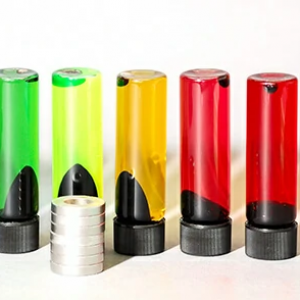
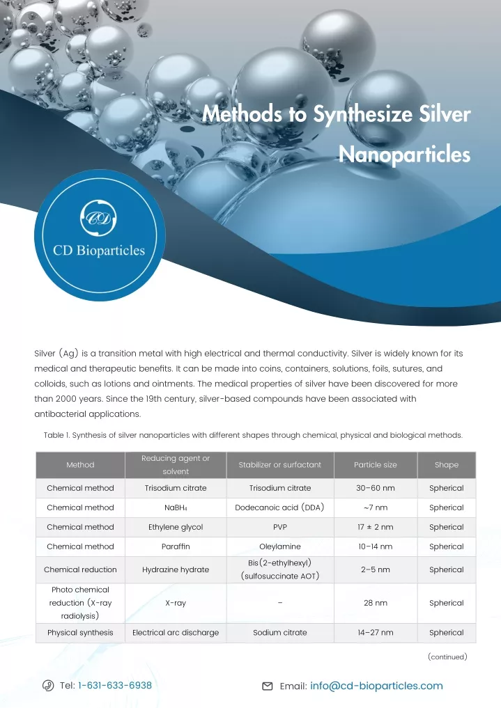
Methods to Synthesize Silver Nanoparticles CD Silver (Ag) is a transition metal with high electrical and thermal conductivity. Silver is widely known for its medical and therapeutic benefits. It can be made into coins, containers, solutions, foils, sutures, and colloids, such as lotions and ointments. The medical properties of silver have been discovered for more than 2000 years. Since the 19th century, silver-based compounds have been associated with antibacterial applications. Table 1. Synthesis of silver nanoparticles with different shapes through chemical, physical and biological methods. Reducing agent or solvent Method Stabilizer or surfactant Particle size Shape Chemical method Trisodium citrate Trisodium citrate 30–60 nm Spherical Chemical method NaBH4 Dodecanoic acid (DDA) ∼7 nm Spherical Chemical method Ethylene glycol PVP 17 ± 2 nm Spherical Chemical method Paraffin Oleylamine 10–14 nm Spherical Bis(2-ethylhexyl) (sulfosuccinate AOT) Chemical reduction Hydrazine hydrate 2–5 nm Spherical Photo chemical reduction (X-ray radiolysis) X-ray – 28 nm Spherical Physical synthesis Electrical arc discharge Sodium citrate 14–27 nm Spherical (continued) Email: info@cd-bioparticles.com Tel: 1-631-633-6938
Table 1. (continued) Physical synthesis TX-100, UV TX-100 30 nm Spherical Biological synthesis Bacillus sp. Bacillus sp. 5–15 nm Spherical Biological synthesis Lactobacillus Lactobacillus Proteins 6–15.7 nm Spherical Shewanella oneidensis Shewanella oneidensis Biological synthesis 2–11 nm Spherical Trichoderma viride Biological synthesis Fungus T. viride 5–40 nm Spherical Cassia angustifolia Cassia angustifolia Biological synthesis 9–31 nm Spherical Daucus carota Daucus carota Biological synthesis 20 nm Spherical Biological synthesis Bacillus strain CS 11 Bacillus strain CS 11 42–92 nm Spherical Aspergillus niger Aspergillus niger Biological synthesis 1–20 nm Spherical Arbutus unedo leaf extract Arbutus unedo leaf extract Biological synthesis 3–20 nm Spherical Chemical method Ethylene glycol PVP – Cubic Chemical method Pentanediol (H-1.5 PDO) PVP – Cubic Chemical method Ethylene glycol PVP 30–50 nm Cubic Carboxymethylated chitosan (CMCTS) Leaf extracts from Eucalyptus macrocarpa Sodium borohydride in the presence of sodium citrate Carboxymethylated chitosan (CMCTS) Leaf extracts from Eucalyptus macrocarpa Photochemical 2–8 nm Cubic 10–50 nm (mean crystallite size = 38±2 nm) Biological synthesis Cubic Wet-chemical – 4 ± 2 nm Nanorods Chemical method Potassium tartaric PVP – Nanorods Chemical method (soft, solution- phase) Diameters of 30– 40 nm Ethylene glycol – Nanowires In diameter 30– 40 nm Wet chemical Ascorbic acid – Nanowires Microwave technique Ethylene glycol PVP – Nanowires Chemical method (polyol) Ethylene glycol PVP – Nanobars Chemical reduction Hydrazine hydrate PVP 50–200 nm Triangular Ethylene glycol monoalkyl ethers Microwave-assisted PVP – Nanoprisms Email: info@cd-bioparticles.com Tel: 1-631-633-6938
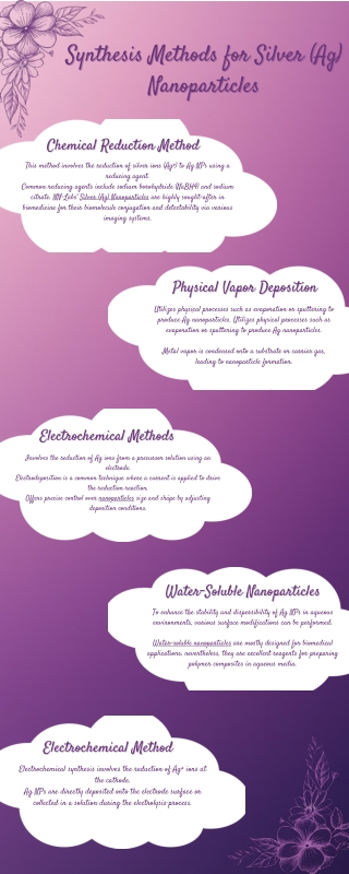
In biology and biomedical research, silver nanoparticles (AgNPs) are important due to their physical and chemical properties. Silver products have strong inhibitory and bactericidal effects and have been used for centuries to prevent and care for various diseases, especially infections. AgNPs are considered to have antifungal, anti-inflammatory, anti-viral, and anti-platelet activities. AgNPs have been considered for a variety of physical, biological, and pharmaceutical purposes. They can be designed to have different forms, including particles, rods, squares, lines, films, and coatings (Table 1). Synthetic methods can be divided into physical, chemical, and biological methods (Figure 1). Advantages and disadvantages of each are shown in Table 2. Figure 1. Diverse synthesis routes of silver nanoparticles. Table 2. Synthesis methods of silver nanoparticle and the corresponding advantages and disadvantages Synthesis methods Advantages Disadvantages Absence of chemical reagents and by-products; Size uniformity High energy consumption; Time-consuming for thermal stability; Surface structural defects Physical methods Controllable silver content; Narrow size distribution Chemical methods Employ of highly deleterious organic solvents Facile and mild; Large-scale; Eco-friendly; Cost effective; Biocompatible; Long-term stable Ambiguous action mechanism; Pretreatment of organic matter; Potential bacterial contamination and transfection Biological methods Email: info@cd-bioparticles.com Tel: 1-631-633-6938
Physical methods Table 3. Synthesis of silver nanoparticles by means of physical methods. Method Shape Silver size (nm) Laser ablation Spherical 31 Laser ablation Spherical 12–29 Laser ablation Irregular 27–41 Laser ablation Spherical 27–120 Laser ablation Spherical 6.48 Laser ablation Spherical 4–18 Laser ablation Spherical 5–13 Laser ablation Spherical 20–51 Laser ablation Irregular 15–20 Laser ablation Spherical 7.9–16.2 Laser ablation Spherical 2.5–8.5 Laser ablation Spherical 10.6±2.6 Laser ablation Spherical 9–15 Laser ablation Spherical 50 Laser ablation Spherical 5–50 Small ceramic heater Spherical 6–21.5 Thermal decomposition Spherical 9.5± 0.7 Thermal decomposition Spherical 14.4±3.3 Thermal decomposition Spherical 4–7 Thermal decomposition Spherical 4.7 Thermal decomposition Spherical 8.0±1.3 Thermal decomposition Spherical 40–50 Email: info@cd-bioparticles.com Tel: 1-631-633-6938
Evaporation–condensation Evaporation-condensation is the physical method for synthesizing AgNPs. It is usually carried out using a tube furnace at atmospheric pressure that is reliable for synthesizing various sizes of nanoparticles. However, the tube furnace takes up a lot of space, consumes a lot of energy while increasing the ambient temperature around the source material, and requires a lot of time to achieve thermal stability. A typical tube heating furnace requires more than a few kilowatts of power consumption and a warm-up time of around ten minutes to reach a stable working temperature. • Laser ablation In another study, AgNPs were synthesized using laser ablation in solution. During the preparation process, the AgNPs were washed with distilled water and placed in a quartz cell filled with 5 mL of high-pressure liquid chromatography (HPLC) grade water. The study was performed using a laser with a repetition frequency of 10 Hz and a pulse width of 5-9 ns. The results show that the average AgNPs size is 12 nm under the action of 532 nm laser, and that prepared under 1064 nm laser is 31 nm. This study concluded that the particle size distribution depends on the laser wavelength. The change in particle size is affected by the laser light absorbed by the particles. Laser ablation is a green technology that can produce stable AgNPs in a variety of dispersion media without using metal precursors and reducing agents. The produced colloid has high purity and unique surface characteristics without any by-products. In principle, these characteristics make AgNPs produced by this method one of the best candidates for antimicrobial applications. However, productivity has not been sufficient for direct use in the industrial sector. • Thermal decomposition In another study, Lee and Kang synthesized AgNPs by thermal decomposition. Their method involves an aqueous solution of AgNO3 and sodium oleate. Spherical AgNPs with an average particle size of 9.5±0.7 nm can be obtained by reacting at room temperature to 290°C for 1h under a slow heating rate of 2°C/min. • Chemical methods Chemical reduction Chemical reduction is the most used method to synthesize AgNPs using organic and inorganic reducing agents (Table 4). This is because the metal surface has free electrons in the conduction band and positively charged nuclei, which continues to produce a colored silver solution through a single process. Long-lasting silver clusters can be formed, confirming the synthesis of AgNPs. Generally, different reducing agents such as sodium citrate, ascorbate, sodium borohydride (NaBH4), elemental hydrogen, polyol process, N,N-dimethylformamide (DMF), ascorbic acid, poly(ethylene glycol)-block copolymers, hydrazine, and ammonium formate can be used to reduce silver ions (Ag+) in aqueous or non-aqueous solutions by one-pot method. • Tel: 1-631-633-6938 Email: info@cd-bioparticles.com
Table 4. Synthesis of silver nanoparticles using chemical reduction. Silver salt Reduction agent Stabilizer/capping agent Silver size (nm) AgNO3 Aniline Etyltrimethlyammonium bromide 10–30 AgNO3 D(+)-Glucose and NaOH - 8 and 24 AgNO3 D-Glucose Carboxy methyl cellulose, NaOH 5–15 AgNO3 Ethylene glycol Poly(vinyl pyrrolidone) 50–175 AgNO3 Ethylene glycol Poly(vinyl pyrrolidone) 8–10 AgNO3 Ethylene glycol Poly(vinyl pyrrolidone) 17±2 AgNO3 Ethylene glycol - 17–70 AgNO3 Gallic acid Gallic acid 7–89 AgNO3 Glucose Poly(vinyl pyrrolidone) 20–80 AgNO3 Hydrazine hydrate and citrate of sodium Sodium dodecyl sulfate 10–20 AgNO3 Hydrazine hydrate and sodium citrate Sodium dodecyl sulfate 10–20 AgNO3 NaOH Alkali lignin (low sulfonate) 5–100 AgNO3 Poly(ethylene glycol) Poly(ethylene glycol) 15–30 AgNO3 Poly(ethylene glycol) - 10–80 AgNO3 Poly(vinyl pyrrolidone) and gelatin Glucose, fructose, lactose, and sucrose 35 AgNO3 Sodium borohydride Tri-sodium citrate ~5 AgNO3 Sodium borohydride - 3.5–6 Microemulsion techniques Microemulsion technology has excellent properties such as low interfacial tension, large surface area, good thermodynamic stability, and the ability to dissolve incompatible liquids. The microemulsion method is flexible that it can organically organize the particle size control mechanism, geometry, morphology, uniformity, and specific surface area. A microemulsion containing silver ions is mixed with another microemulsion containing a reducing agent, as shown in Figure 2. The collision and coalescence of the droplets lead to the reduction of silver ions in the water core and produce silver precipitation. • Email: info@cd-bioparticles.com Tel: 1-631-633-6938
Figure 2. Schematic of the synthesis of AgNPs with water-in-oil (w/o) microemulsion. Microwave-assisted synthesis Contrary to traditional heating techniques, microwave synthesis uses variable-rate microwave radiation to reduce AgNPs. This technology gives up a faster reaction and provides a higher AgNPs concentration under the same temperature and exposure conditions. Zhao . developed a simple, environmental friendly, and efficient method for synthesizing uniform spherical AgNPs. Sodium alginate is used as a stabilizer and reducing agent to synthesize AgNPs in an aqueous medium treated by microwaves. The AgNPs prepared by this method are homogeneous and stable in solution at room temperature (25°C) for 6 months without any signs of agglomeration. • et al Biological methods In recent years, the biological synthesis has received more attention. It designed to minimize the negative impact on the environment. The use of chemical methods to synthesize AgNPs requires three main components: silver salt, reducing agent, and stabilizer, or capping agent. In biological methods, reducing agents and stabilizers are replaced with molecules obtained from organisms such as plants, bacteria, fungi, yeast, and algae. Using plant extracts Plant extracts can be used as reducing agents to synthesize AgNPs and provide an environmental friendly alternative. As a naturally occurring resource, its cost is low, and the resources are abundant. Table 5 shows several plant extracts used to synthesize AgNPs from their leaves, seeds, roots, and fruits. Song . proposed a biological method using leaf extracts of five plants such as Pine, Persimmon, Ginkgo, Magnolia, and Platanus as reducing agents. The results show that the reaction temperature, leaf fluid concentration and AgNO3 can control the size of AgNP. Studies have shown that from the perspective of synthesis rate and conversion rate, the magnolia leaf is the best reducing agent for the synthesis of AgNPs. This method can synthesize AgNPs with an average particle size of 15-500 nm. • et al Email: info@cd-bioparticles.com Tel: 1-631-633-6938
Table 5. Plant-mediated synthesis of silver nanoparticles. No. Plant Average size (nm) 1 Barley,flax, ryegrass 0.6–2 Lycopersicon esculentum mill, Piper pedicellatum, Centella asiatica l., Azadirachta indica leaf, Triphala 30–40, 2–3, 30–50, 43 and 59 respectively 2 3 Mangosteen leaf extract 50 Tragia involucrata, Cymbopogon citronella, Solanum verbascifolium and Tylophora ovata leaf extracts Clitoria ternatea and Solanum nigrum leaf extracts 4 32, 36, 41 and 28 respectively 5 10–50 Cinnamomum tamala leaf extract 6 10 Murraya koenigii leaf extract 7 10–25 Azadirachta indica leaf extract 8 34 Ananas comosus leaf extract 9 12 M. balbisiana, Azadirachta indica and O. tenuiflorum 10 200 Seaweed Gracilaria birdiae 11 20.3–94.9 12 Pepper leaf broth 5–60 Desmodium plant 13 10 Rumexhymenose palus root extracts 14 2–40 Catharanthus roseus leaf extract 15 35–55 Using fungus as reduction agents In addition to plant extracts, fungi can also be used to synthesize AgNPs (Table 6). Mukherjee et al. recommend synthesizing AgNPs using the fungus Verticillium. The study found that the average particle size of AgNP is 25±12 nm. In addition, Fusarium oxysporum was also used as a biological reducing agent to synthesize AgNPs. 10-3 M silver nitrate was mixed with 10g of Fusariumoxysporumin a conical flask filled with 100mL of distilled water. This method can be used to prepare spherical and occasionally triangular AgNPs with a particle size ranging from 5 to 15 nm. • Email: info@cd-bioparticles.com Tel: 1-631-633-6938
Table 6. Fungus-mediated synthesis of silver nanoparticles. Fungus Size (nm) Shape Location Aspergillu flavus 8.92 Spherical Cell wall A. fumigatus - - Extracellular A. terreus 1–20 Spherical Extracellular Cladosporium cladosporioides 10–100 - - Coriolus versicolor 25–75, 444–491 Spherical Extracellular and intracellular Humicola sp. 5–25 Spherical Extracellular Macrophomina phaseolina 5–40 Spherical Cell-free filtrate P. fellutanum 5–25 Spherical Extracellular P. nalgiovense AJ12 25 ± 2.8 Spherical Cell-free filtrate P. sajor-caju 30.5 ± 4.0 Spherical Extracellular Trichoderma asperellum 13–18 Nanocrystalline Extracellular T. viride 5–40 Spherical Extracellular T. viride 2–5 Spherical Cell free extract • Using microorganisms as reduction agents In recent years, microorganisms such as bacteria (Table 7) and yeast have been considered as alternative reducing agents for the rapid AgNPs synthesis. Shahverdiet al. proposed using the culture supernatants of Klebsiella pneumonia, Escherichia coli, and Enterobacter cloacae. The results show that AgNPs can be synthesized within 5 minutes, with an average particle size of 52.5 nm. Recently, Shanthi et al. used a probiotic strain B.licheniformis Dahb1 as a silver nitrate-reducing agent to synthesize AgNPs. Placing the probiotic in a flask with sterile nutrient broth and incubating at 37°C for 24 hours can obtain spherical AgNP with a particle size of 18-63 nm. Table 6 summarizes the various microorganisms used to synthesize AgNPs. Email: info@cd-bioparticles.com Tel: 1-631-633-6938
Table 7. Bacteria-mediated synthesis of silver nanoparticles. Bacteria Size (nm) Shape Location Acinetobacter calcoaceticus 8–12 Spherical Extracellular A. haemolyticus MMC8 4–40 - Extracellular Enterobacter aerogenes 25–35 Spherical Extracellular Escherichia coli 42.2–89.6 Spherical Extracellular Klebsiella pneumoniae 15–37 Spherical Extracellular Morganella sp. 10–40 Quasispherical Extracellular Proteus mirabilis 10–20 Spherical Extracellular and intracellular Pseudomonas aeruginosa SM1 6.3±4.9 Spherical, disk-shaped Extracellular Vibrio alginolyticus 50–100 Spherical Extracellular and intracellular Xanthomonas oryzae 14.86 Spherical, triangular, rod-shaped Extracellular B. flexus 12 and 65 Spherical and triangular Extracellular Exiguobacterium sp. 5–50 Spherical Extracellular Ureibacillus thermosphaericus 10–100 Spherical Extracellular In summary, physical, chemical, and biological methods are used to synthesize AgNPs. Physical methods have the disadvantages of large space requirements, high energy consumption, and a long time to achieve thermal stability. The chemical method can produce AgNPs easily, but the toxicity of its by-products is a major issue. Biological methods are getting popular because of their environmental friendliness and low cost. Creative Diagnostics provides a comprehensive list of silver nanoparticles with different surface properties in multiple sizes. Silver nanoparticles produced with our proprietary protocols are highly monodisperse with a narrow size distribution. Please visit our website to see more. Email: info@cd-bioparticles.com Tel: 1-631-633-6938
References: 1. Lee, S. H., & Jun, B. H. (2019). Silver Nanoparticles: Synthesis and application for nanomedicine. International journal of molecular sciences, 20(4), 865. 2. Tsuji, T., Iryo, K., Nishimura, Y., & Tsuji, M. (2001). Preparation of metal colloids by a laser ablation technique in solution: influence of laser wavelength on the ablation efficiency (II). Journal of Photochemistry and Photobiology A: Chemistry, 145(3), 201-207. 3. Lee, D. K., & Kang, Y. S. (2004). Synthesis of silver nanocrystallites by a new thermal decomposition method and their characterization. Etri Journal, 26(3), 252-256. 4. Zhao, X., Xia, Y., Li, Q., Ma, X., Quan, F., Geng, C., & Han, Z. (2014). Microwave-assisted synthesis of silver nanoparticles using sodium alginate and their antibacterial activity. Colloids and Surfaces A: Physicochemical and Engineering Aspects, 444, 180-188. 5. Song, J. Y., & Kim, B. S. (2009). Rapid biological synthesis of silver nanoparticles using plant leaf extracts. Bioprocess and biosystems engineering, 32(1), 79. 6. Mukherjee, P., Ahmad, A., Mandal, D., Senapati, S., Sainkar, S. R., Khan, M. I., ... & Sastry, M. (2001). Fungus-mediated synthesis of silver nanoparticles and their immobilization in the mycelial matrix: a novel biological approach to nanoparticle synthesis. Nano letters, 1(10), 515-519. 7. Ahmad, A., Mukherjee, P., Senapati, S., Mandal, D., Khan, M. I., Kumar, R., & Sastry, M. (2003). Extracellular biosynthesis of silver nanoparticles using the fungus Fusarium oxysporum. Colloids and surfaces B: Biointerfaces, 28(4), 313-318. 8. Shahverdi, H. R., Jamalifar, H., & Nohi, A. A. (2007). Biomimetic synthesis and characterization of protin capped silver nanoparticles. Pro. Biochem, 42, 919. 9. Shanthi, S., Jayaseelan, B. D., Velusamy, P., Vijayakumar, S., Chih, C. T., & Vaseeharan, B. (2016). Biosynthesis of silver nanoparticles using a probiotic Bacillus licheniformis Dahb1 and their antibiofilm activity and toxicity effects in Ceriodaphnia cornuta. Microbial pathogenesis, 93, 70-77. 10. Solanki, J. N., & Murthy, Z. V. P. (2011). Controlled size silver nanoparticles synthesis with water-in-oil microemulsion method: a topical review. Industrial & engineering chemistry research, 50(22), 12311-12323. For more information, view our website: www.cd-bioparticles.com Email: info@cd-bioparticles.com Tel: 1-631-633-6938 Fax: 1-631-938-8221 Address: 45-1 Ramsey Road, Shirley, NY 11967, USA 5