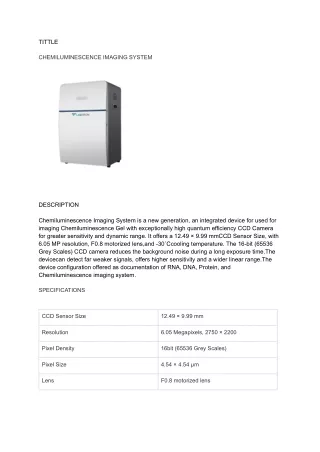
Download
1 / 8
0 likes | 6 Views
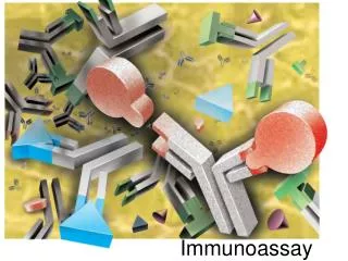
Overview of Magnetic Beads-based Chemiluminescence Immunoassay

E N D
Overview of Magnetic Beads-based Chemiluminescence Immunoassay CD Immunology was established in the 18th century with the study of microbiology, and entered the development period from the 19th to the middle of the 20th century. During this period, the general understanding of the immune function in the scientific society has changed due to the shift from observing human phenomena to conducting experiments. The period of modern immunology began from the middle of the 20th century. Immunoassay is based on the specific reaction between antigen and antibody, and the detection signal is amplified and displayed by isotopes, enzymes, chemiluminescence substances, and so on. It is often used to detect proteins, hormones, and other trace substances. The nature and development of various immunological methods are detailed in Table 1. Immune diagnosis plays a very important role in clinical diagnosis. Table 1. Properties and characteristics of various immune detection techniques RIA 10-15 >15% 105 3-4h + ELISA 10-13 10%-15% 102 2-3h - Manual/batch- automatic 6-12M Qualitative/semi -quantitative CLIA 10-15-10-18 <10%-15% 106-7 65min - Manual/fully automatic 12M ECLIA 10-17 5% 107 10min - Magnetic Beads-based CLIA 10-21 <10% 107 18-40min - Sensitivity Between-run precision Linearity and range Time cost Radiation pollution Operation Manual Fully automatic Fully automatic Shelf life 2M 12M 12M Qualitative/quantitative Quantitative Quantitative Quantitative Quantitative Tel: 1-631-633-6938 Email: info@cd-bioparticles.com
Chemiluminescent immunoassay (CLIA) was developed in the late 1970s. The principle of this technique is to use the free energy released by the chemical reaction to excite the intermediate from the excited state to the ground state (commonly used alkaline phosphatase-amantadine amine, horseradish peroxidase-luminol derivatives, horseradish peroxidase-luminol derivatives). During this process, equal-level photons are released, and the quantitative analysis is accomplished by measuring the photons (Figure 1). CLIA is a very excellent quantitative analysis method. It has the specificity of fluorescence; at the same time, it does not need the excitation light, avoiding its stray light from interfering with the analysis and improving the sensitivity. Moreover, CLIA avoids environmental pollution and health hazards caused by radiation analyses. Energy Substrate (ground state) Optical Signal Enzyme Catalysis Substrate (excited state) Solid-phase Figure 1. Schematic of the basic principle of chemiluminescence. Characteristics and Development Trend of CLIA CLIA is a highly sensitive technique. All antigenic substances (including semi-antigens) can be determined by CLIA. CLIA began to show the following characteristics in the 1990s when it was developed rapidly: (1) highly sensitive, with a limit of 10-17 to 10-21 mol / L, which is much higher than that of radioimmunoassay (RIA) and enzyme-linked immunosorbent assay (ELISA). Its sensitivity is similar to the time-resolved fluorescence immunoassay (TRFIA) but the cost is lower. (2) strong specificity and good reproducibility (CV < 10%). (3) determination range is wide, up to 7 orders of magnitude. (4) its reagent has good stability and pollution-free period of 12 months. (5) easy to operate and can be automated. Tel: 1-631-633-6938 Email: info@cd-bioparticles.com
Magnetic beads-based CLIA is an already established and relatively easy technique that combines magnetic separation technology, chemiluminescence technology, and immunoassay technology (Figure 2). This technique makes the full use of the rapid and easy automation of magnetic separation technology, the high sensitivity of chemiluminescence technology, and the specificity of immunoassay. It is irreplaceable in the field of biological analysis. In order to further improve the detection sensitivity of chemiluminescence, using nanoparticles to immobilize enzyme-labeled antibodies can increase the number of enzyme-labeled antibodies bound to the surface of a single antigen, thus amplifying the signals. Luminescent Substrate Magnetic Beads Figure 2. Schematic of the principle of magnetic beads-based CLIA (double antibody sandwich CLIA). Magnetic Beads for CLIA Types of magnetic beads for CLIA By material • Material Advantages Disadvantages There are many gaps in the silica structure, which can easily cause a large amount of water accumulation on the surface, increasing the complexity of chemical reaction. Very unstable under alkaline conditions There is a large amount of non-specific adsorption Relatively strong mechanical strength Better chemical stability Magnetic silica beads Good hydrophilicity Good blood compatibility Copolymerization with other polymer compounds can improve its mechanical properties and stability Magnetic PAM/PAA beads Excessive shrinkage High mechanical strength Surface easy functionalization Weak non-ionic surface interaction Cross-linked polystyrene can maintain a stable structure in strong acids and bases Controllable particle size The surface of polystyrene microspheres is hydrophobic, which is non-specific to some proteins, so the surface needs to be functionalized Magnetic polystyrene beads Tel: 1-631-633-6938 Email: info@cd-bioparticles.com
By functional groups • Common Activation Methods Reaction Site Hydroxyl magnetic beads CDI, cyanogen bromide activation Amino group Carboxyl magnetic beads EDC, NHS activation Amino group Amine magnetic beads Glutaraldehyde activation Amino group Epoxy magnetic beads - Sulfhydryl, amino group NHS-activated magnetic beads - Amino group Streptavidin magnetic beads Biotin labled ligand Biotin Selecting proper magnetic beads for CLIA Magnetic beads used in CLIA require rapid magnetic response, monodisperse, uniform particle size, good suspension, low non-specific adsorption, and good stability. When selecting beads, the following aspects should be considered. Magnetic beads for CLIA have a wide size range. It is best to try different magnetic bead sizes to determine the best size that will suit your experiment and bring you the best results. The gold standard of the bead size is about 1.0 µm. You can start from this to speed up your optimization. Size of the beads Small magnetic beads may have advantages over large ones, but their magnetic separation speed is slower. Understand the properties of each material and determine how each material affects your assay. Bead material Polystyrene and polystyrene-divinylbenzene are two commonly used materials for the magnetic beads. We recommend the bead density to be greater than 1.2g/cc. The higher the percentage of magnetic pigment (30-50% recommended), the faster the separation of magnetic beads. Density of the beads Higher density also means a faster sedimentation rate. The plain surface allows the passive coupling of your antibody. But it also can have specificity problems and high background readings. The modified surface, such as -COOH group, -NH2 group, -OH group or -SH group modification, allows the covalently coupling of the antibodies to the beads to obtain a more stable coating. Aggregation could be a potential problem. Surface properties Pre-activated surfaces, such as those containing tosyl, chloromethyl, or epoxy groups, allows the stable covalent coupling of your antibody/antigen. The cost may be high. Bio-activated surfaces, such as those activated with streptavidin, anti-IgG, or protein A or G, allows the attachment of the antibodies via bioconjugation technology. Therefore, only the biomagnetic separation is needed to separate free antibodies or antigens from those that react with each other and the centrifugation step can be skipped. However, biotin or streptavidin beads are expensive. Starting from a small-scale test to determine the type of the beads to use will save a lot of trouble later when you decide to scale up your assay production. Batch size Small batches must be verified separately, while large batches can be verified quickly. Tel: 1-631-633-6938 Email: info@cd-bioparticles.com
According to our experience, here are a few popular combinations for magnetic beads-based CLIA. Beads Size Surface modification Magnetic pigment (by weight) 700 nm-1.2 µm Carboxyl 35-65% 0.9-1.4 µm Carboxyl 15-50% (Encapsulated) 1-2 µm Amino 35-65% 900 nm-1.8 µm Amino 35-50% (Encapsulated) 1.1-1.4 µm Tosyl >30% Tel: 1-631-633-6938 Email: info@cd-bioparticles.com
Basic Principles of CLIA Indirect method • The indirect method is to coat the antigen on the magnetic beads, and then use the enzyme-labeled anti-antibodies to detect the antibodies in the samples (Figure 3), which is suitable for detecting antibody samples. This method is relatively convenient. As long as the coating antigen is changed, different analytes can be detected. However, the specific antibodies in the markers will competitively bind to antigens, resulting in false-negative results. Enzyme-labeled anti-antibody Antigen Antibody as analyte Mag Figure 3. Basic principle of chemiluminescence detection using indirect method. Capture method • Specific IgM and specific IgG for certain antigens are often present in serum at the same time, and the latter will interfere with the detection of IgM antibodies. Therefore, the capture method is often used to determine IgM antibodies. (Figure 4). All serum IgM (including specific IgM and non-specific IgM) are immobilized on the solid phase, and the specific IgM is determined after IgG is removed. Specific antigen Antibody Enzyme-labeled antibody Mag IgM Figure 4. Basic principle of chemiluminescence detection using capture method. Solid-phase antigen competitive method • The competitive method refers to the competitive binding of tested antibodies and enzyme-labeled antibodies to the antigen on the surface of the magnetic beads (Figure 5). The amount of enzyme-labeled antibody bound to the solid phase carrier is inversely proportional to the amount of antibody in the sample. If there is no antibody in the tested sample, the enzyme-labeled antibody can bind to the antigen smoothly and show a strong signal. If there are antibodies, the competitive binding between the enzyme-labeled antibody and the antibody in the sample to the antigen will weaken the binding power of the enzyme-labeled antibody to the antigen, as well as the signal intensity. Tel: 1-631-633-6938 Email: info@cd-bioparticles.com
Antibody as analyte Antigen Enzyme-labeled antibody Mag Figure 5. Basic principle of chemiluminescence detection using competitive method. Sandwich method • Sandwich CLIA is a detection method combining double antibody sandwich method with chemiluminescence method (Figure 6). First, the microtiter plate is pre-coated with specific antibodies conjugated magnetic beads against the analyte. Then, the standard or sample is added to the microplate wells, and the analyte in the standard and sample will bind to the immobilized antibody. Next, add the biotin-conjugated antibody, which will bind to the analyte adsorbed on the plate. The complex of two antibodies and analyte in the well acts as a "sandwich" structure. After washing away the free biotin-labeled antibody, adding avidin-labeled horseradish peroxidase (HRP) to each microtiter plate, and incubation, luminol is added for quantitative analysis. Enzyme-labeled secondary antibody Antibody Antigen as analyte Mag Figure 6. Basic principle of chemiluminescence detection using sandwich method. Procedures for Developing Magnetic Beads-based CLIA When developing and using CLIA, it is important to follow the basic procedure as shown below to ensure that your assay is effective and accurate. Define the expected dynamic range (e.g. the approximate concentration range and optimal concentration range of your analyte). Define the specimen to be tested (e.g. blood, serum, urine, etc.). Select the antibodies to be used in your assay (e.g. pAb for not well-defined epitopes, or mAb for well-defined epitope and has very high specificity). Choose the most suited magnetic beads (e.g. size, surface modification, etc.). Optimize the coating procedure (e.g. adsorption properties of beads, coupling strategy, etc.). Select the homogenization method (e.g. roller homogenization, overhead homogenization, vortexing or • • • • • • Tel: 1-631-633-6938 Email: info@cd-bioparticles.com
Conclusion CLIA has the advantages of strong specificity, high sensitivity, high accuracy, simple and rapid operation. It combines the advantages of chemical analysis and immunological analysis. It has been widely used in life science, environmental science, clinical medicine, physiology, and other fields. Magnetic beads-based CLIA is gradually replacing the existing chemiluminescence method and becoming the standard method in antigen and antibody immunoassay. For more information, view our website: www.cd-bioparticles.com Email: info@cd-bioparticles.com Tel: 1-631-633-6938 Address: 45-1 Ramsey Road, Shirley, NY 11967, USA Fax: 1-631-938-8221 5