Download
1 / 65
650 likes | 655 Views
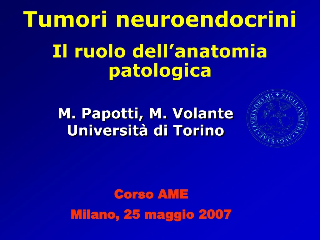
Tumori neuroendocrini Il ruolo dell’anatomia patologica. M. Papotti, M. Volante Università di Torino. Corso AME Milano, 25 maggio 2007. Caro Mauro, ….

E N D
Tumori neuroendocrini Il ruolo dell’anatomia patologica M. Papotti, M. VolanteUniversità di Torino Corso AME Milano, 25 maggio 2007
Caro Mauro, …. Riguardo alla tua relazione, considerato il titolo, avremmo piacere che tu affrontassi nel tempo che hai a disposizione 20 min la classificazione del neuroendocrino, i limiti della citologia in rapporto alle scelte terapeutiche, il discorso dei recettori e della loro utilità in pratica clinica. considera che sarà un corso AME (…) con un taglio decisamente pratico e questo anche per quanto riguarda l'anatomia patologica. Invito del dr R. Cozzi
Different diagnostic criteria and classification for • Medullary thyroid carcinoma • Pheochromocytoma/Paraganglioma • Parathyroid adenoma/carcinoma • Pituitary adenoma/carcinoma
neuroendocrine tumor classificationMENU - History, Definition & Glossary - Pathological classification pure NE tumors mixed NE/exocrine tumors - Further characterisation
History 1907 “carcinoid” term Oberndorfer 1930 carcinoid syndrome Cassidy 1940/50 “helle zellen” Feyrter 1952 serotonin discovery Erspamer & Asero 1963 fore-, mid-, hind-gut carcinoids Williams & Sandler 1965/70 APUD concept Pearse 1980 diffuse NE system WHO 1994 “neuroendocrine tumor” replaces carcinoid Capella et al 2000 classification of endocrine tumors WHO Solcia et al
History 1907 “Karzinoid” term SiegfriedOberndorfer 6 ileal tumors, slowly growing, no metastases
NE TUMORS Glossary & Synonyms • carcinoid, malignant carcinoid • apudoma • islet cell tumor • adenoma / microadenoma vs carcinoma • Kultchisky cell tumor / carcinoma • endocrine carcinoma • endocrine neoplasm • hormone…-oma (insulinoma, gastrinoma,..) • pancreatic (A/B/D/PP)-cell tumor
Neuroendocrine tumor classificationMENU - History, Definition & Glossary -Pathological classification pure NE tumors mixed NE/exocrine tumors - Further characterisation
THE BENIGN THE MALIGNANT THE CLINICALLY AGGRESSIVE Clinical Morphological Hormonal Ki67 Gene expression WHICH CLASSIFICATION CRITERION ????
Clinical Morphological Hormonal Ki67 Gene expression Incidence, Localisation, Endocrine function, Symptoms, Treatment, Behavior, Survival
Clinical Morphological Hormonal Ki67 Gene expression insulinoma Macro 1 cm • Single or multiple • Well demarcated or invasive • Size 0.5-15 cm. • Cystic appearance • Lymph node mets • Distant mets Gastric micro-carcinoids
Micro Tumor architecture
Clinical Morphological Hormonal Ki67 Gene expression THE BENIGN THE MALIGNANT THE CLINICALLY AGGRESSIVE Gross features and structure are not enough for the purpose of identifying homogeneous groups with clinical relevance
CLASSIFICATION PROBLEMS 1994 2000
2000 WHO CLASSIFICATION • Introduces terms tumor/carcinoma (vs carcinoid) ….LUNG EXCLUDED • Replaces neuroendocrine with endocrine • Incorporates hormonally active tumors • (morpho-functional approach) • Recognizes mixed exo-endocrine tumors • Incorporates information on tumor grade • Introduces criteria of malignancy (angioinvasion, Ki67 pancreas, only)
Microscopic features of malignancy • Depth of invasion • Size of tumor • Angioinvasion • High grade cellular atypia GI NETS
PANCREATIC NETs Microscopic features of malignancy • Invasion of surrounding tissues • Size of tumor (>6 cm is suspected) • High grade cellular atypia • Focal or diffuse necrosis • High mitotic index • Blood or lymph vessel & nerve invasion • Ki67 > 2%
Does this system fit with the clinical practice? Is it easily applicable? E. Bajetta, L. Catena, G. Procopio, E. Bichisao, L. Ferrari, S. Della Torre, S. De Dosso, S. Iacobelli, R. Buzzoni, L. Mariani and J. Rosai Is the new WHO classification of neuroendocrine tumours useful for selecting an appropriate treatment? Ann Oncol 2005 16:1374 Problem of other non-carcinoid NETs (eg malignant pheochromocytoma, MTC, parathyroid carcinoma, Merkel cell carcinoma, etc…)
Does this system fit with the clinical practice? Is it easily applicable? • Benign vs uncertain behaviour in well differentiated pancreatic NETs • Appendiceal NETs (small & invasive?) • Mixed endocrine – exocrine tumors • Use morphology or Ki67 first? • The new TNM system for foregut NETs • Extra GEP: pulmonary NETs especially intermediate grades
BENIGN vs UNCERTAIN BEHAVIOR Benign Uncertain Extrapancreatic growth no no Angioinvasion no yes Size >=2 cm no yes Mitoses >2 /10HPF no yes Ki67 >2% no yes
Resembles WD NET (benign), but in general: size >3 cm, mitoses >2, Ki67 >5% butabove all: unequivocal signs of malignancy must be present LOCAL INVASION LYMPHNODE LIVER
FOREGUT NET CLASSIFICATION Recent proposal of a staging system for fore-gut NETs and improvement of grading system using mitoses + Ki67
SPECTRUM OF PULMONARY NETS TC SCLC Difficulties in identifying the intermediate entities AC LCNEC
SPECTRUM OF PULMONARY NETS TC AC SCLC Indolent clinical behavior Aggressive clinical behavior Significantly different survival Arrigoni et al 1972
SPECTRUM OF PULMONARY NETS TC AC LCNEC SCLC <2 mitoses 3-10 mitoses >10 mitoses small cells no necrosis or necrosis (necrosis) (necrosis) Significantly different survival p<0.0001 Significantly different survival p<0.0001 NO significantly different survival Travis et al Am J Surg Pathol 22,934,1998
SPECTRUM OF PULMONARY NETS TC AC LCNEC SCLC TC AC LCNEC SCLC NO significantly different survival Asamura et al JCO 24:70,2006
Degree of differentiation WELL DIFF. POORLY DIFF. LUNG SMALL CELL/ LARGE CELL NE CARCINOMA TYPICAL CARCINOID ATYPICAL CARCINOID MEEC WELL DIFF. NE TUMOR benign/borderline WELL DIFF. NE CARCINOMA POORLY DIFF. NE CARCINOMA (small/large cell) GEP LOW GRADE HIGH GRADE Biological behaviour
Neuroendocrine tumor classificationMENU - History, Definition & Glossary -Pathological classification pure NE tumors mixed NE/exocrine tumors - Further characterisation
2000 WHO CLASSIFICATION CATEGORIES RECOGNIZED • 1 well differentiated endocrine tumor • 2 well differentiated endocrine carcinoma • 3 poorly differentiated endocrine carcinoma • 4 mixed exocrine-endocrine tumor • (5 tumor-like lesions)
CGA Exocrine Endocrine
NE differentiation in colon cancer 1995 CGA mucin
…..ANY BIOLOGICAL and/or CLINICAL SIGNIFICANCE?? pure non-NE ca. non-NE ca. focal NE BREAST and COLORECTAL CANCERS NO Non-NE carcinomas with NE differentiation Y/N ? NSCLC STOMACH & PROSTATE CANCERS YES Breast (WHO2004) Lung (WHO2004) GI tract (WHO2003) Prostate (WHO2003)
Am J Surg Pathol August 2006 LCNEC of the stomach are significantly more aggressive than conventional ADC. ADC-NED have also a worse prognosis than conventional ADC (borderline significance)
Reduced survival of CGA+ cases CGA increase during progression
CASE REPORT April 2007 July 1996 Nov 1997 Oct 1999 May 2000 Rectal polyp, cancerised Local recurrence Pelvic recurrence Stable disease Rectum resection Radical Surgery FNAB ADC stage B1 • ChTx 1: 5-FU + folic acid (6x) • RT: 50 Grey • ChTx 2: oxaliplatin + 5-FU + folic acid (12x) CgA CgA CgA Oncologia Prof Dogliotti dr Tampellini ChTx 1 RT ChTx 2
pure NE tum. pure non-NE ca. NE tum. non-NE ca. mixed (intermingled) mixed (collision) focal non-NE focal NE 0% 30% 100% non-NE NE 100% 30% 0% Mixed NE/non-NE carcinomas WDET - TC WDEC - AC PDEC – SCLC/LCNEC Non-NE carcinomas with NE differentiation Breast (WHO2004) Lung (WHO2004) GI tract (WHO2003) Prostate (WHO2003) WHO 2000 Endocrine WHO 2004 lung WHO 2000 Endocrine tumors
Degree of differentiation WELL DIFF. POORLY DIFF. LUNG SMALL CELL/ LARGE CELL NE CARCINOMA TYPICAL CARCINOID ATYPICAL CARCINOID Mixed Endo-Exocrine Ca WELL DIFF. NE TUMOR benign/borderline WELL DIFF. NE CARCINOMA POORLY DIFF. NE CARCINOMA (small/large cell) GEP LOW GRADE HIGH GRADE Biological behaviour
HOW TO BREAK THE ARROW? TYPICAL CARCINOID LUNG LUNG WELL DIFF. NE TUMOR benign/borderline ATYPICAL CARCINOID GEP MEEC SMALL CELL/ LARGE CELL NE CARCINOMA WELL DIFF. NE CARCINOMA POORLY DIFF. NE CARCINOMA (small/large cell) Light microscopy + ……?? GEP
Neuroendocrine tumor classificationMENU - History, Definition & Glossary - Pathological classification pure NE tumors mixed NE/exocrine tumors - Further characterisation
Further characterisation • hormonal • Ki 67 • receptors • molecular THE BENIGN THE MALIGNANT THE CLINICALLY AGGRESSIVE
Chromogranins Synaptophysin N-CAM Cytoker. 34bE12 NSE, VMAT PGP9.5 Neurofilaments MARKERS SSTR2 Hormones: calcitonin, bombesin, insulin, glucagon, somatostatin, gastrin, PP, VIP, ACTH, serotonin, … NEW: ghrelin, cortistatin, obestatin, secretagogin[Virch Arch 449, 402, Oct 2006] CGA
Further characterisation • Ki 67 proliferation index LOW HIGH
KI-67: DIAGNOSTIC USE 2000
KI-67: PROGNOSTIC USE OS KI-67 in WD NE carcinoma P = 0.017 Prognostic value if groups are homogeneous (eg all WDCA or PDCA). Otherwise possible bias due to tumor differentiation TTP P = 0.043 Brizzi et al, 2007 (submitted)