Download
1 / 38
390 likes | 547 Views
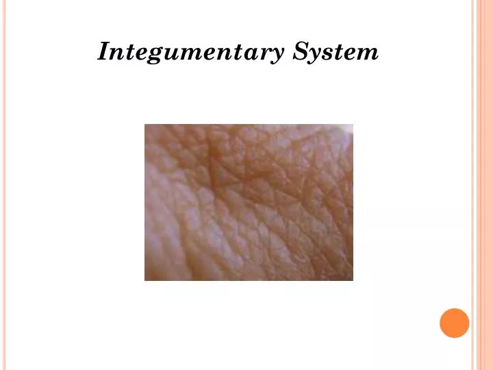
Integumentary System. Introduction. The integumentary system protects the body from environmental damage. The skin forms a protective barrier shielding the body from the elements and pathogens, as well as performing several other vital functions.

E N D
Introduction • The integumentary system protects the body from environmental damage. • The skin forms a protective barrier shielding the body from the elements and pathogens, as well as performing several other vital functions. • Skin is essential to well-being, helps to regulate body temperature, and contains many accessory organs such as nails, hair, and glands.
System Overview • The integumentary system is comprised of the skin and its accessory components including hair, nails, and associated glands.
System Overview • The integumentary system performs several vital functions: • Protection from pathogens • Balances fluid levels • Stores fatty tissue for energy supply • Produces vitamin D (with help from the sun) • Provides sensory input • Helps to regulate body temperature
The Skin • The skin is the largest organ, weighing approximately 20 pounds and covering an area about 20.83 square feet on an adult. • A cross section of skin reveals three layers: • Epidermis • Dermis • Subcutaneous fascia
Epidermis • The epidermis is the layer of skin that we see on the outside. It is made up of five even smaller layers of tissue. • There are no blood vessels in this layer. • The cells on the surface of the epidermis are constantly shedding, being replaced with new cells that grow and arise from the deeper region called the stratum basale every 2–4 weeks.
Epidermis • The outermost layer is a layer of dead cells, called the stratum corneum, which are flat, scaly, keratinized epithelial cells. • You slough off 500 million cells every day, or about 1½ pounds of dead skin a year, allowing for rapid repair in case of injuries. • Keratinization- epithelial cells lose their moisture and are replaced by horny tissue horny tissue= tough fibrous layer of keratin keratin= fibrous structural protein
Skin Color • Specialized cells called melanocytes are located deep in the epidermis and are responsible for skin color. • Melanocytes produce melanin, a substance that causes skin color. • Variation in skin color is the result of the amount of melanin produced and how it is distributed, not the number of melanocytes. • Carotene, another form of pigment, gives a yellowish hue to skin while a pinkish hue is derived from the hemoglobin in the blood.
Effects of Disease on Skin Color • Color of skin can indicate disease. • When liver disease occurs, the body can’t break down bilirubin. The buildup of bilirubin gives the skin a deeper, yellow color. • A malfunctioning adrenal gland can cause the skin to turn bronze due to excessive melanin.
Effects of Disease on Skin Color • Excessive bruising could indicate skin, blood, or circulatory problems. • Cyanosis, or a blue coloring, results from a drop in oxygenation.
Dermis • The layer below, or inferior, to the epidermis is the thicker dermis layer. • This layer contains the following: • Capillaries • Collagenous/elastic fibers • Involuntary muscles • Nerve endings • Lymph vessels • Hair follicles • Sudoriferous glands (sweat) • Sebaceous glands (oil)
Dermis • Small “fingers” of tissue project from the surface of the dermis and anchor this layer to the epidermal layer. • Nerve fibers allow you to sense what is happening in your environment. • Vasodilation of capillaries in this layer causes blushing.
Dermis • Collagen and elastic fibers allow for the elasticity of skin, preventing the tearing of skin with movement. They allow skin to return to normal shape during periods of rest. Older people lose some of this elasticity, leading to wrinkles. Small fingerlike projections
Sudiferous Glands • Two main types of sudiferous, or sweat, glands • Apocrine sweat glands secrete at the hair follicles in the groin and anal region as well as the armpits and become active around puberty and are believed to act as sexual attractants. • Eccrine glands are found in greater numbers on your palms, feet, forehead, and upper lip and are important in the regulation of temperature.
Sudiferous Glands • The body has 3 million sweat glands. • Sweat has no odor, but bacteria degrades the substances in the sweat over time into chemicals that give off strong smells commonly known as body odors.
Sebaceous Glands • Sebaceous glands play an important role by secreting oil, or sebum. • Sebum keeps the skin from drying out and (due to its acidic nature) helps destroy some pathogens on the skin’s surface.
Subcutaneous Fascia • The innermost layer of the skin is the subcutaneous fascia, or hypodermis. • The subcutaneous fascia is composed of elastic and fibrous connective tissue and fatty tissue. • Lipocytes, or fat cells, produce the fat needed to provide padding to protect the deeper tissues of the body and act as insulation for temperature regulation. • Fascia attaches to the muscles of the body.
Fascial tissue makes up the primary unit of structure in the human body. Fascia is a three dimensional, web like, network of connective tissue which wraps all of the muscles, bones, organs, and brain.
How Skin Heals • If skin is punctured and the wound damages blood vessels, the wound fills with blood. • Blood contains substances that cause clotting. • The top part of the clot exposed to air hardens to form a scab, nature’s band-aid, forming a barrier and preventing pathogens from entering. • In minor wounds, the dermis will eventually regenerate. In severe wounds, the dermis will be replaced by a scar
Burns to the Skin • Burns can be caused by heat, chemicals, electricity, or radiation. • Two factors affect assessments of damage: • Depth • Amount of area damaged • The depth of a burn relates to the layer or layers of skin affected by the burn
First Degree Burns • First degree burns damage only the outer layer, or epidermis. • Symptoms include redness and pain, but no blister. • Pain subsides in 2-3 days and there is no scarring. • Complete healing takes about 1 week.
Second Degree Burns • Second degree burns involve the entire depth of the epidermis and a portion of the dermis. • Symptoms include redness, pain, and blistering. • The extent of blistering is dependent on the depth of the burn.
Second Degree Burns • Blistering extends after the initial burn. • Blisters heal within 10-14 days if there are no complications, with deeper second degree burns taking 1-3½ months. • Scarring in second degree burns is common.
Third Degree Burns • Third degree burns affect all three layers of the skin. • The surface of the burn has a leathery feel and will range in color from black, brown, tan, red, or white. • The victim feels no pain because the pain receptors are destroyed. • Also destroyed are sweat and sebaceous glands, hair follicles, and blood vessels
Fourth Degree Burns • Fourth degree burns are the worst burns. • These burns penetrate the bone and cause bone damage.
Amount of Area Damaged • The rule of nines is used to estimate the extent of area damaged by burns. • The body is divided into the following regions, each given a percentage of body surface area value: • Head and neck – 9% • Each upper limb – 9% (2 x 9 = 18%) • Front of trunk – 18% • Back of trunk and buttocks – 18% • Front of legs – 18% • Back of legs – 18% • Perineum (including anus and urogenital region) – 1%
Burns – Clinical Concerns • The clinical concerns for burn victims relate to the functions of the skin already discussed, including: • Bacterial infections • Fluid loss • Heat loss
Burn Treatment • Severe burns require healing steps at an intensity level the body can’t manage on its own. • Damaged skin must be removed as soon as possible and skin grafting must be started. • Autografting is using the patient’s own skin, while heterografting (from a donor) is required if the patient suffered a large area of burn and has little healthy skin to graft.
Burn Treatment • Grafting requires many trips to the OR because large areas can’t be done all at once and often the grafts don’t “take.” • It is possible to grow sheets of skin tissue in the laboratory from patient cells or utilization of synthetic materials. • Grafting requires many trips to the OR because large areas can’t be done all at once and often the grafts don’t “take.” • It is possible to grow sheets of skin tissue in the laboratory from patient cells or utilization of synthetic materials.
Coming Attractions NAILS HAIR TEMPERATURECONTROL DISEASES