Download
1 / 23
230 likes | 239 Views
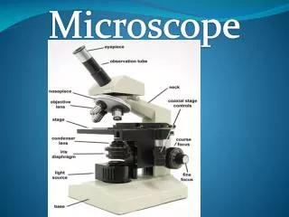
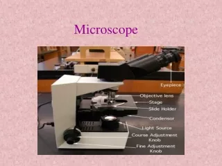
Microscope. Basics. T. Trimpe 2005 http://sciencespot.net/. Base. - Single piece of metal -Support here when moving. Arm. -Hold here when moving. Stage. - Supports specimen -Hole allows light to pass through. Diaphragm. - controls amount of light -ranges 1-5. Stage Clips.

E N D
Microscope Basics T. Trimpe 2005 http://sciencespot.net/
Base -Single piece of metal -Support here when moving
Arm -Hold here when moving
Stage -Supports specimen -Hole allows light to pass through
Diaphragm -controls amount of light -ranges 1-5
Stage Clips -secures specimen/slide
Bulb/Mirror -Light source -NEVER use the sunlight it will hurt your eyes!!
Scanning Objective Lens -Shortest lens • 4x magnification
Low Power Objective Lens • Medium length lens • 10x magnification
High Power Objective Lens • -Longest lens • 40x magnification
Body Tube -Allows light from objective to pass upward
Eye Piece/Ocular lens -Site of observation • 10x magnification
Nose Piece -Allows you to switch objective lenses
Coarse Adjustment Knob -Used to focus specimen -DO NOT use on high power objective lens
Fine Adjustment Knob -Used for minute focusing -Used after coarse adjustment knob
Total magnification = (ocular lens power) x (objective lens power) • Example: (10x)ocular lens x (40x)high power lens = 400x total magnification • Magnification: The ability to increase the size of an image X = 400x total magnification Objective Lens Ocular Lens
We can see better details with higher the powers of magnification, but we cannot see as much of the image. Which of these images would be viewed at a higher power of magnification? Comparing Powers of Magnification
Light Pathway: Light Source → Specimen → Objective Lens → Body Tube → Eye Piece
Resolution- add this to your notes! • making the image clear • ability to determine between two objects or points • can be adjusted with the fine/course knob or the diaphragm (to allow more light in)
Use of a Scope • Carry the microscope with 2 hands (arm & base) • Looking at a slide: • 1. Use ONLY ONE slide at a time • 2. ALWAYS start at LOW power first(10x) • 3. ALWAYS start with stage lowered completely • 4. Make sure the objective clicks into position • 5. The coarse focus knob is ONLY used on the lowest power lens • 6. The fine focus knob is used on all lenses to bring the image into focus
Putting the Microscope Away • Lower the stage completely and click the lowest objective into place • Return slide to tray • Wind up the scope power cord
2. Electron microscope: Uses electrons instead of light to visualize the specimen, electrons bounce off or pass through and a computer interprets a picture Always black and white, specimen must be dead to examine Scanning EM – magnifies up to 100,000x Transmission EM – magnifies up to 200,000x Types of Microscopes • 1. Compound Light Microscope: Shines light through a specimen and uses 2 lenses for magnification • Magnification – increase in an objects apparent size • Resolution – how clear the image appears once magnified • Max clear magnification ~2000x
Microscope Images Compound Light Microscope - Protozoa Scanning Electron Microscope (SEM) Butterfly Tongue Transmission Electron Microscope - Bacteria