Download

1 / 33
330 likes | 343 Views
MESODERMAL DERIVATIVES. By: Dr. Mujahid Khan. Derivatives. Connective tissue Cartilage Bone Striated & smooth muscles Heart Blood & lymphatic vessels Kidneys, ovaries, testes & genital ducts Serous membrane lining the body cavities Spleen & cortex of the supra renal gland.

E N D
MESODERMAL DERIVATIVES By: Dr. Mujahid Khan
Derivatives • Connective tissue • Cartilage • Bone • Striated & smooth muscles • Heart • Blood & lymphatic vessels • Kidneys, ovaries, testes & genital ducts • Serous membrane lining the body cavities • Spleen & cortex of the supra renal gland
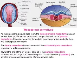
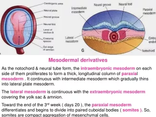
Development of Somites • As the notochord and neural tube forms • Embryonic mesoderm on each side of them proliferates • Form a thick longitudinal columns of paraxial mesoderm • Each column is continuous with intermediate mesoderm
Development of Somites • Intermediate mesoderm gradually thins into a layer of lateral mesoderm • Lateral mesoderm is continuous with the extraembryonic mesoderm • Extraembryonic mesoderm covers the yolk sac and amnion
Somites • Paraxial mesoderm differentiates and begins to divide into cuboidal bodies called somites by the end of 3rd week • These blocks of mesoderm are located on each side of developing neural tube • About 38 pairs of somites form during the somite period of human development (20-30 days)
Somites • About 42-44 pairs of somites are present by the end of 5th week • Are triangular in transverse section • Form distinct surface elevations on the embryo • Are used as one of the criteria to know the age of the embryo at this stage
Somites • First appear in the future occipital region • Soon develop craniocaudally • Gives rise to the axial skeleton and associated musculature • Also forms adjacent dermis of the skin • The first pair of somites appear at the end of 3rd week
Somites • First appear at a short distance caudal to the cranial end of the notochord • Subsequent pairs form in a craniocaudal sequence
Intraembryonic Coelom • Also known as primordium of embryonic body cavity • Appears as isolated coelomic spaces in the lateral mesoderm and cardiogenic mesoderm • These spaces soon coalesce to form a single horseshoe shaped cavity called intraembryonic coelom
Parietal & Visceral Layers • Somatic or parietal layer continuous with the extraembryonic mesoderm covering the amnion • Splanchnic or visceral layer continuous with the extraembryonic mesoderm covering the yolk sac
Parietal & Visceral Layers • Somatic mesoderm with overlying embryonic ectoderm form the embryonic body wall or somatopleure • Splanchnic mesoderm with underlying embryonic endoderm form the embryonic gut or splanchnopleure
Fate of Intraembryonic Coelom During the 2nd month, the intraembryonic coelom is divided into 3 body cavities: • Pericardial cavity • Pleural cavity • Peritoneal cavity
Early Development of Cardiovascular System • Starts at the beginning of the 3rd week • Vasculogenesis and angiogenesis begins in the extraembryonic mesoderm of the yolk sac, connecting stalk and chorion • Embryonic blood vessels begin to develop about 2 days later
Early Development of Cardiovascular System • The urgent need for blood vessels to bring nourishment and oxygen to the embryo from mother causes the early formation of the cardiovascular system • A primordial uteroplacental circulation develops during the 3rd week • Until then the embryonic nutrition is obtained from maternal blood by diffusion
Vasculogenesis & angiogenesis Formation of embryonic vascular system involves 2 processes: • Vasculogenesis • Angiogenesis
Vasculogenesis • Mesenchymal cells differentiate into endothelial precursors called Angioblast • Angioblast aggregate to form isolated angiogenic cell clusters or blood islands • Small cavities appear within the blood islands • Angioblasts flatten to form endothelial cells
Vasculogenesis • Endothelial cells arrange themselves around the cavities in blood island to form the endothelium • These endothelium lined cavities soon fuse to form networks of endothelial channels called Vasculogenesis
Angiogenesis • Vessels sprout into adjacent areas by endothelial budding and fuse with other vessels called Angiogenesis
Development of Blood Cells • Blood cells develop from the endothelial cells of vessels called hemangioblasts • Develop at the end of 3rd week on the yolk sac and allantois • Hematogenesis does not begin until 5th week • It occurs first in liver and later in spleen, bone marrow & lymph nodes
Development of Blood Cells • Fetal and adult erythrocytes are derived from different hematopoietic progenitor cells (hemangioblasts) • Mesenchymal cells surrounding the primordial endothelial blood vessels differentiate into the muscular and connective tissue elements of the vessels
Primordial Cardiovascular System • Heart & great vessels develop from mesenchymal cells in the cardiogenic area • Paired longitudinal endothelial lined channels or endocardial heart tubes develop during the 3rd week • These tubes fuse to form the heart tube
Primordial Cardiovascular System • The tubular heart joins with blood vessels in the embryo, connecting stalk, chorion and yolk sac to form a primordial cardiovascular system • Heart begins to beat on 21-22 days and blood circulates • CVS is the first organ system to reach a functional state
Further Development of Chorionic Villi • Primary chorionic villi becomes secondary chorionic villi as they acquire mesenchymal cores • Before the end of third week capillaries develop in the secondary chorionic villi • Now it is called tertiary chorionic villi
Further Development of Chorionic Villi • Cytotrophoblastic extensions from these stem villi join to form a cytotrophoblastic shell that anchors the chorionic sac to the endometrium • The rapid development of chorionic villi during the third week greatly increases the surface area of chorion • This causes exchange of oxygen and nutrients between the maternal and embryonic circulations