Download
1 / 42
440 likes | 500 Views
Purulent Meningitis in Children. Jiang Li Department of Neurology Children’s Hospital Chongqing University of Medical Sciences. Purulent Meningitis. Acute infection of central nervous system(CNS). 90% of cases occur in the age of 1mo-5yr.

E N D
Purulent Meningitis in Children Jiang Li Department of Neurology Children’s Hospital Chongqing University of Medical Sciences
Purulent Meningitis • Acute infection of central nervous system(CNS). 90% of cases occur in the age of 1mo-5yr. • The inflammation of meninges caused by various bacteria.Common features in clinical practices include: fever, increased intracranial pressure, meningeal irritation. One of the most potentially serious infections, associated with high mortality (about 10%) and morbidity.
Etiology • 1.1 Pathogens: • Main pathogens: Neissria meningitidis, streptoccus pneumoniae, Haemophilus influenzae. (2/3 of purulent meningitis are caused by these pathogens) • Pathogens in special populations (neonate & <3mo infants , malnutrition, immunodeficiency): gramnegative enteric bacilli, group B streptococci, staphlococcus aureus
1.2 Major risk factors for meningitis • Immature immunologic function and attenuated immunologic response to pathogens Low level of immunoglobulin, defects of complement and properdin system Immature or impaired blood-brain-barrier (BBB) Immature BBB function: maturation at about 1yr Impaired BBB: Congenial or acquired defects across mucocutaneous barrier
1.3 Access of bacteria invasion Typical access---hematogenous dissemination Bacteria colonizing the mucous membranes of the nasopharynx invasion into local tissue bacteremia hematogenous seeding to the subarachnoid space Mode of transmission: Person to person contact through respiratory tract secretions or droplets
Access of bacteria invasion • Bacteria spread to the meninges directly: through anatomic defects in the skull or head trauma • Invasion from parameningeal organs: such as paranasal sinuses or middle ear
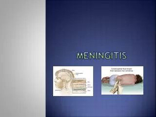
2. Pathology • Structure of meninges
Pathology • Characterized by leptomeningeal and perivascular infiltration with polymorphonuclear leukocytes and an inflammatory exudate. • Exudate which may be distributed from convexity of brain to basal region of cranium. • Exudate is more thickness due to streptococcus pneumoniae than other pathogens.
3. Clinical manifestations • The younger the child is, the higher incidence of meningitis will be. ½-2/3 of cases occur less than 1yr of age. • Mode of presentation: Acute or fulminant onset: symptoms and signs of sepsis; meningitis evolve rapidly over a few hours and death within 24 hours; usually infected with Neissria meningitides (N. meningitides).
Mode of presentation • Subacute onset: Precede by several days of upper respiratory tract or gastrointestinal symptoms; difficult to pinpoint the exact onset of meningitis; usually with meningitis due to Haemophilus influenzae (H influenzae) and streptoccus pneumococcus (S pneumococcus).
Clinical manifestations • Common features of meningitis: signs of systemic infection : fever(90-95%), anorexia,shock, alteration of mental status and consciousness neurological signs: increased intracranial pressure: headache, vomiting(82%), herniation meningeal irritation: nuchal rigidity(77%), kernig sign, brudzinski sign
Clinical manifestations • Seizure (20-30%) Focal or generalized Due to cerebritis, infarction, electrolyte disturbances Frequently noted with H influenzae & S pneumococcal meningitis Persist after 4th day and difficult to treat with poor prognosis
Clinical manifestations • Alteration of mental status and consciousness Including: irritability, lethargy, stupor obtundation, coma Due to increased intracranial pressure, cerebritis, hypotension Often with pneumococcal or meningococcal meningitis Comatose patients with a poor prognosis
Clinical manifestations • The symptoms and signs are not evident in neonates and infants younger than 3mo of age; and patients already received irregular antibiotic therapy.
Clinical manifestations Comparison of the manifestations of meningitis between different age groups
4. Diagnosis • Earlier diagnosis and prompt initiation of effective antibiotic treatment is critical for minimizing sequelae of purulent meningitis. Suspected cases: febrile infants with seizure, meningeal irritability, increased intracranial pressure, altered mental status Pay attention to the atypical symptoms and signs in neonate, infant and patient already received irregular antibiotic therapy
Diagnosis • Diagnosis is confirmed by analysis of cerebrospinal fluid ( CSF) Suggestion bacterial meningitis Increased pressure (90%) Appearance: slightly cloudy to purulent Raised white blood cells,consisting chiefly of polymorphonuclear leukocytes Raised protein concentration, decreased glucose concentration (80%)
Diagnosis • Confirmation of the diagnosis: isolation from the CSF of a specific bacterial pathogen by microscopy or a positive culture or rapid antigen- detection test of CSF Gram-stained smear of CSF: identify the causative organism in 70-90% of cases CSF culture: positive in about 80% of cases. definitive diagnosis, determination of antibiotic sensitivity. PCR: amplifies bacterial DNA (H influenzae, N. meningitidis)
5. Differential diagnosis • Purulent meningitis caused by different pathogens Neissria meningitidis: Occur in epidemics (type A,C), which is more common in spring, or sporadic all the year (type B,C,Y) Sudden onset with various cutaneous signs ( petechiae, purpura, or an erythematous macular rash)
Differential diagnosis Streptococcus pneumoniae: Young infants ( <1yr) are most susceptible population Peak season: spring and winter Easier to have subdural effusion and hydrocephalus Easily have a protracted course and relapse
Differential diagnosis • Haemophilus influenzae Occurs predominantly in infants 2mo to 2yr of age Many cases are in winter Higher incidence of subdural effusion • Others pathogens: staphylococcus aureus, gramnegative enteric bacilli Special susceptible population: neonate, <3mo infants, malnutrition, immunodeficiency Severe infection, difficult to treat
Differential diagnosis • Meningitis caused by other microorganisms Viral meningitis/encephalitis: Less severe systemic infectious symptoms Usually not develop after 2-3weeks CSF: normal glucose
Differential diagnosis Tuberculous meningitis Subacute onset and progress A history of close contact with known cases of tuberculosis Evidence of acute or healed tubercular infection on chest x-ray Tuberculin skin test : OT, PPD CSF
6. Complications and sequelae • 6.1 Subdural effusion • Definitive diagnosis: volume of fluid in subdural space >2ml, protein>0.4g/L, • Incidence: develop in 10-30% of patients, asymptomatic in 85-90% of patients; especially common in infants 4-6 month of age ( rare in children over 1yr);
subdural effusion • Causative organisms: 45% of cases of meningitis caused by H influenzae, 30% by S pneumoniae, 9% by N meningitidis
subdural effusion Indications: No response to a sensitive antibiotic therapy Prolonged fever or fever reoccurring after an afebrile interval with effective treatment Bulging fontanel, widening of sutures, enlarging head circumference, emesis,seizure, altered consciousness. Improved CSF profile with more serious clinical manifestations
subdural effusion • Diagnosis methods: Cranial translucent test B ultrasonic examination and CT Subdural space puncture normal subdural effusion
Complications 6.2 Ventriculitis 6.3 hydrocephalus
Complications • 6.2 Ventriculitis • Usually occurs in neonates and infants (<1yr), with severe prognosis • The main cause is delayed diagnosis and treatment of meningitis.
Ventriculitis • Diagnosis: B ultrasonic examination or neuroimaging studies( CT, MRI): enlarged lateral ventricle Lateral ventricle puncture: bacteria and inflammatory cells in ventricular fluid, WBC>50x106/L, Glucose<1.6mmol/L, or protein>400mg/L.
Complications 6.3 hydrocephalus : Communicating hydrocephalus: adhered or destroyed arachnoid granulation around the cistern at the base of the brain Obstructive hydrocephalus: following obstructed of the cerebral aqueduct, or the foramina of Magendie and Luschka 6.4 others: Deafness, blindness, paralysis, epilepsy, mental retardation
Treatment • 7.1 Antibacterial therapy • Therapy principles: early treatment, antibiotics susceptible to pathogens and with high permeability through BBB, given intraveninously, enough dose, enough course of antibiotic therapy
Antibacterial therapy Susceptible to pathogens First choice: Cefotaxime, Ceftriaxone (3dr generation of cephalosporins, high permeability through BBB, products of metabolism also has effect, CSF sterilization within 24h) Other choice: Penicillin, Chloromycin, Cefuroxime, Ceftazidime ( delayed effect to make CSF sterile, high incidence of relapse and deafness)
Treatment 7.2 Supportive care • Maintenance fluid and thermal energy supplement: Fluid administration: 60-80ml/kg/day Fluid infusion with dehydration therapy
Treatment • increased intracranial pressure • Osmotic therapy: intravenous mannitol 0.5- • 1g/kg/every time, q4-6h • Combination with intravenous dexamethasone: • 0.3-0.5mg/kg/day • Endotracheal intubation and hyperventilation
Treatment • Subdural effusion Few volume could be absorbed with treatment spontaneously Subdural puncture: take out 15ml/each time (unilateral puncture), less than 30ml/each time ( bilateral puncture), everyday or every other day Stripping operation: for the cases not cure after 3-4weeks
Treatment • Others: Ventriculitis : lateral ventricle puncture and injection of antibiotics locally Epilepsy: AEDs