Download
1 / 50
560 likes | 777 Views
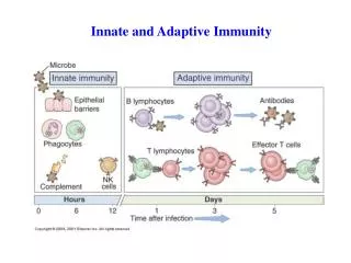
The dark side of innate and adaptive immunity - cytotoxic and dendritic cell lymphomas. …attack of the clones. Dominic Spagnolo. PathCentre WA. Dr Vincent McGovern 1915-1983. 2005. The players. Components of the innate and adaptive arms of the immune system

E N D
The dark side of innate and adaptive immunity - cytotoxic and dendritic cell lymphomas …attack of the clones Dominic Spagnolo PathCentre WA Dr Vincent McGovern 1915-1983 2005
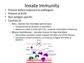
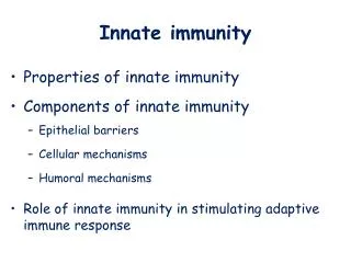
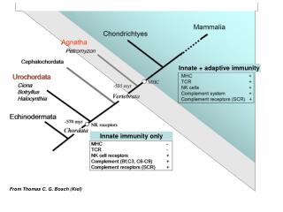
The players • Components of the innate and adaptive arms of the immune system • extreme form of immune dysregulation • CD8+ (ab+) T cells • gd+ T-cells • NK cells • NK-associated T-cells • Dendritic cell (type 2) precursor
Why?? • Follicular lymphomas are boring • Very different from B-cell lymphomas • least common of the uncommon (<2%) • diagnostically problematic (mimic other NHL) • phenotypic promiscuity • diagnostic armamentarium • clinically distinctive • extranodal, limited stage • tissue restricted lymphocyte subsets • aggressive, chemoresistant • hemophagocytic syndrome • chronic antigenic stimulation, impaired immunity
Agenda 1. Review clinicopathological features • WHO 2001 • WHO-EORTC (2005) classification for cutaneous lymphomas 2. Normal lymphocyte immunobiology 3. New data 4. CD4+ CD56+ hematodermic neoplasm
immature DC Mature DC Myeloid progenitor CD4+ MARROW STEM ab+ T cell L2 L3 90-95% CD8+ Common lymphoid progenitor gd+ T cell 5-10% B cell NK
DENDRITIC CELLS NK CELLS Macrophages, pmn, mast cells INNATE immunity B-1 B cells gd +T CELLS NKT CELLS Treg cells INNATE-LIKE “Bridge” innate and adaptive B cells, plasma cells ab + T CELLS - CD8+ - CD4+ ADAPTIVE immunity 10 20 lymphoid tissues Blood Tissue-restricted - Specific tissue distributions - - Site-specific homing mechanisms -
Natural killer lymphocytes NK and NK/T lymphomas
NCR CLR CD56 NK CELL perf TCR - G granz CD3e TIA-1 KIR costimulatory Natural cytotoxicity non-MHC cytotoxicity Immunoregulatory cytokines chemokines (innate immunity) (adaptive immunity) Phenotypic promiscuity!
Natural killer receptors (NKRs) NEJM 2000; 343:37-49
NK receptors Most NK receptors are not specific for NK cells activating or inhibitory activating • HLA-recognising receptors • Killer Ig-like receptors (KIR) • C-type lectin-like receptors (CLR) • CD94/NKG2 family heterodimers • Non-HLA binding • NKG2D homodimer • Natural cytotoxicity receptors (NCR) • NK-specific: 2 NCRs only (NKp46; NKp30) • Costimulatory receptors • e.g. 2B4; NTBA (activ/inhib)
Diagnostic utility of NKR expression • Abnormal patterns of NKR expression • restricted repertoires (KIR) indicative of pathological state, not necessarily lymphoma • receptor genes do not rearrange • transcripts may be of prognostic relevance • normal reference ranges to be established • correlation with other clonality studies
NK cell proliferations WHO E/nodal NK/T-cell lymphoma, nasal type Aggressive NK cell leukemia Chronic NK-lymphocytosis
E/nodal NK/T-cell lymphoma, nasal type • Asia, C & S America; American Indians • rare in West • Upper respiratory tract - nasal, n/pharynx • rhinitis, bleeding, mass • bone destruction - high frequency • maxilla, orbit, palate • Skin, subcutis, GIT, lung, testis, female genit. • skin second most common site • Extranasal cases - more often advanced stage
Extranodal NK/T lymphomas • Morphology heterogeneous, clinically irrelevant • Necrosis, apoptosis • death receptor activation: Fas-FasL, TRAIL --> --> caspase cascade --> apoptosis • cytotoxic granule exocytosis • angio-invasion, angio- destruction • chemokines Mig and IP-10 endotheliotoxic (CXC family) • Activated cytotoxic phenotype • Perforin+, Granzyme+ (cf. TIA-1+ only) • EBER+ (site-, geography-dependent)
Extranodal NK/T-cell lymphoma, nasal type • NKR expression • CD94/NKG2A+ majority • CD94 mRNA +ve ?? better prognosis than -ve • KIR+ minority of cases; restricted repertoire • Cutaneous lymphocyte antigen+ (>50%) • cutaneous cases particularly • poor prognostic factor • Del (6q); gains at 1p32-pter • multiple recurrent gains/losses
E/nodal NK/T-cell lymphoma, nasal type • Aggressive, even stage 1E • early systemic spread, typically extranodal • skin, subcutis, GIT, testis, soft tissue; marrow • leukemic (aggressive NK cell leukemia) • chemoresistance - MDR-1 (P-glycoprotein) • hemophagocytic syndrome ~10% (TNFa; NFkB) • 5 yr OS and DFS <40%; DXT mainstay of Rx • IPI of prognostic significance • no effect - age, stage, p53, cell size, EBER, Rx, lineage (Ng SB et al; Mod Pathol 2004)
BLOOD 1996; 87:1474-1483 BLOOD 1997; 89:4501-4513 EXTRANODAL - liver, spleen, intestine, lung, URT Little or no peripheral adenopathy Immunosuppressed CD56 POSITIVITY Male predominance Advanced stage, aggressive
Aggressive NK-cell leukemia • Japan, Asia >>> West (RARE!) • largest series 22 cases (Suzuki R, Leukemia 2004) • Relatively young, fever, B symptoms, anemia • Leukemic presentation • minority lymphoma-like • adenopathy, little or no blood involvement • Hepatosplenomegaly; coagulopathy • frequent GIT, skin, nodes; marrow subtle or overt • large granular lymphocytosis • Aggressive - death in months; HPS frequent
Aggressive NK-cell leukemia • sCD3-, cCD3+, CD4-8-, CD56+ • TCR germline • EBER+ • SKY and CGH studies • frequent 6q, 13q, 11q, 17p13 deletions (similar to NK/T lymphoma, nasal type) • translocations involving Xp21-pter; 8p23 Message: • Don’t undercall as an indolent large granular lymphocytosis / leukemia
The mucosal immune system CD3 Enteropathy type T-cell lymphoma
Enteropathy-type T-cell lymphoma(ETTL) • Arises from phenotypically heterogeneous subsets of IELs • cytotoxic T lymphocytes • natural killer cells (rare) • Coeliac disease - clinical or latent • HLA DQA1*0501; DQB*0201; DRB*0304 • adults, recent onset • refractory sprue • ulcerative jejunitis
Mucosal lymphocyte trafficking thoracic duct mesent LN 4 EPI CCL 25 5 5 3 IEL aEb7 [CD103; HML-1] CCR9 Peyer’s patch T 1 Ag M 2 E- Cad IEL lamina propria 6 MAdCAM-1 L-selectin a4b7 integrin
NORMAL COELIAC REFRACTORY SPRUE / UJ ETTL TCRab CD3 MAJORITY RARE MAJORITY ‘Type A’ EPI 70% CD8 TCRab IEL CD103 3- cCD3+ TCRgd UP TO 30% MAJORITY 8- CD3 M TCR- (or gd+) sCD3-, cCD3+ CD4-, CD8- CD103+ (8+) 15% CD8aa CD103 IEL MINORITY ‘Type B’ CD103 RARE TCRgd TCRgd 3- CD3 CLONAL TCRg-R “Cryptic ETTL” cCD3+ 15% CD8aa CD8 aa CD56 CD56 UNCOMMON
Enteropathy-type T-cell lymphoma • Localised disease typically • may disseminate • LN, liver, spleen, lung, skin • Mass may be absent • ulcers, obstruct, perforate, bleed • Enteropathic mucosa ~50% • proximal >> distal; affected by gluten free diet • Aggressive course - death in months • Not all intestinal T-NHL are ETTL !!
Enteropathy-type T-cell lymphoma • CGH and microsatellite studies • >85% recurrent gains/losses at several loci • gains at 9q33-34 ~60%, both types A and B • C-ABL and NOTCH-1 • >3 imbalances prognostically unfavorable • LOH at 9p21 one third cases (type A >> B) • site of suppressors p14/ARF, p15/INK4b, p16/INK4a
Cutaneous cytotoxic T-cell lymphoma (non-ALCL) • Similar homing mechanisms as in intestine • Skin-homing T cells express: • CLA: binds to E-selectin on cutaneous vascular endothelium • CCR4: binds to CCL17(TARC) expressed on cutaneous vascular endothelium • CCR10: ligates CCL27 on keratinocytes
Aggressive epidermotropic CD8+ CTCL Provisional entity WHO/EORTC 2005 Prof. L Cerroni Localised or disseminated Patches, plaques, papulonodules, tumours Ulceration Adnexotropism, fat rimming Extranodalspread - lung, testis, CNS, oral Median survival 32 mos 0% 5 yr survival
Gamma-delta (gd) T-cells gd+ T-cell lymphomas
gd+ T cells • “Innate-like” lymphocytes • bridge innate and adaptive immune systems • rapid cytokine producers; cytotoxicity • important roles in • infection • immune regulation • immune surveillance • Restricted tissue distribution • 5 - 15% of peripheral blood lymphocytes • 10 - 15% splenic red pulp • thymus, nodes, GIT, other mucosae, liver, skin
gd+ T cells • Lack recirculation • Receptors of very limited diversity (Ig-like) • No MHC restriction or Ag processing • No clonal expansion • Not a homogeneous population • most CD4-8-; IELs are CD8aa+ • subclassification according to V segment usage • Vd1 - naïve/fetal T-cell phenotype (CD45RO-) - spleen, thymus, germinal centres of nodes • Vd2 - memory/adult phenotype (CD45RO+) - blood, interfollicular nodes & tonsils, skin
gd+ T-cell lymphomas • Sites • hepatosplenic (immature Vd1+) - WHO 2001 • mucocutaneous and epithelial sites (mature Vd2) • Limited stage, but aggressive • necrosis, apoptosis • hemophagocytic syndrome • Mimic and overlap other cytotoxic lymphomas • SPTCL, NK/T nasal type, ETTL
Hepatosplenic T-cell lymphoma • Most from naïve splenic gd+ T-cells (Vd1+) • Young adult males • hepatosplenomegaly • thrombocytopenia, anemia, leukopenia (~45%) • no adenopathy; other sites rare • leukemic presentation rare • ab+ cases occur (females; wider age range) • Chronic Ag stimulation + altered immune state • post-organ transplant, SLE, HD, chronic Hep B
Hepatosplenic T-cell lymphoma • Marrow+, but may be subtle, sinusoidal • Lymphocytosis minor or absent at Dx • blastic change +/- leukemic terminally • Aggressive course - poor chemosensitivity • median survival 16 months • indolent, relapsing prodrome in some • Must exclude mimics • T-LGL • aggressive NK leukemia • others
Blood 1998; 91: 1723 - 1731 Arnulf, B et al • Skin/subcutis • GIT, lung, URT • Testis, breast, thyroid • Primary nodal- uncommon • Lymphoblastic 50% • T-cell LGLL - uncommon • Clinicopathologically similar to • other cytotoxic lymphomas at • the same sites • Activated cytotoxic phenotype • cf. hepatosplenic
BLOOD 2003; 101:3407-3412 33 cases gd phenotype: independent predictor of decreased survival • Extremities, trunk • Mucosal, extranodal • - nodes, spleen, marrow • uncommon • HPS in SPTCL-like cases • Chemoresistant • - 15 mos median survival • Mature, cytotoxic, Vd2+ • CD4-, CD8- (few CD8+) • CD56+/-; EBER-
Subcutaneous panniculitis-like T-cell lymphoma (WHO-EORTC 2005)
Subcutaneous panniculitis-like T-cell lymphoma • Mainly young adults, indurated s/cutaneous • nodules/plaques, extremities > trunk > face • - extracutaneous spread rare • Systemic symptoms frequent; • haemophagocytic syndrome up to 50% • Aggressive typically • median survival 27 mos (Go & Wester, review 2004) • high dose chemo/stem cell Tx effective in some • some indolent, remitting/relapsing with chemoRx, • related to phenotype • rarely may disseminate to nodes, e/nodal sites
Subcutaneous panniculitis-like T-cell lymphoma (SPTCL) Pre- 2005 view 2 forms of SPTCL • ab+ SPTCL (75%) • gd+ SPTCL (25%)
BLOOD 2005; 105:3768-3785 ab+ SPTCL • WHO-EORTC 2005 ‘SPTCL’ limited to this type • CD8+, CD56- • limited to subcutis • indolent gd+ SPTCL • WHO-EORTC 2005 classified as gd+ CTCL (provisional entity) • CD4-, CD8-, CD56+ • +/- dermal / epidermal • aggressive
WHO-EORTC 2005 CD4+ CD56+ hematodermic neoplasm(Blastic NK-cell lymphoma) DC2 plasmacytoid monocytes Plasmacytoid dendritic cell (pDC) precursor a neoplasm of plasmacytoid dendritic cell precursors
Monocyte DC1 Myeloid progenitor CD14+ CD11c+ TH1 IL12 IL4 GMCSF TLR 2,3-6,8 signalling T CELL ACTIVATION TLR 7,9 signalling IL3 CD40L(CD154) CD34+ progenitor IFNa CD123+ CD14- CD11c- TH2 Lymphoid progenitor Plasmacytoid DC DC2
J Exp Med 1997; 185:1101-1111 Nature Med 1999; 5:919-923
Am J Surg Pathol 1997; 21(10):1223-1230 Am J Surg Pathol 1999; 23(2):137-146
BLOOD 2002 Feuillard J • marrow (80%) • LN (>50%) • spleen/liver (20%) • >60% stage IV at Dx CD4+ CD56+ hematodermic neoplasm(Blastic NK-cell lymphoma) - WHO/EORTC • Rare, aggressive • Cutaneous +/- leukemic • M>F, median 60s • Violaceous skin plaques, nodules, non-ulcerating
CD4+ CD56+ hematodermic neoplasm(Blastic NK-cell lymphoma) - WHO/EORTC • Leukemic - often at Dx, or develops rapidly • Poor prognosis - median survival 13 mos • Indolent course in some ?predictable • possible favorable factors • skin-confined; age <40; TdT+ • Rx with leukemia-type regimes • induction of remission + allogeneic SCT
CD4+ CD56+ hematodermic neoplasm(Blastic NK-cell lymphoma) • Lineage negative (T-, B-, Myeloid, NK-) • CD45RA+; Granz B; TdT -/+ • Antigen receptor genes germline • Cutaneous lymphocyte antigen +ve • BDCA-2, BDCA-4 positive (novel markers) • del(5q)
CD4+ CD56+ hematodermic neoplasm(Blastic NK-cell lymphoma) • Association with MDS or MPD (15-20%) • AMML evolution in small subset • Acquisition of some myeloid markers during disease progression, e.g. CD33 • ?? Relation to myelo-monocytic lineage • plasticity; common ancestry pDC, Myeloid, NK
Malignant lymphoma of plasmacytoid T-cells Morphologic and immunologic studies characterising a special type of T-cell Hans Konrad Muller-Hermelink et al Am J Surg Pathol 1983; 7(8):849-862 (Myelomonocytic leukemia 3 mos after Dx)
Am J Surg Pathol 2004; 28:585-595 CMML - 4 Unclass. chronic MPD - 1 AML - 1 MPD/MDS - 1 AMoL - 2 • pDC accumulations in Nodes, Marrow, Skin • Clonal identity 1 case by FISH • AML and pDC cells both with monosomy 7 • Most CD56 negative
PathCentre Michael Platten, Lorella Manso Jeremy Taylor, Suzanne Cairns ACKNOWLEDGEMENTS King Edward Memorial Hospital Fremantle Hospital Princess Margaret Hospital Western Diagnostic Pathology St. John of God Pathology Royal Perth Hospital