Download
1 / 25
250 likes | 516 Views
Optimizing Usage of Microfluidic Chemotaxis Chambers for Cancer Research. Kathleen O’Hara Virginia Polytechnic Institute and State University Biological Sciences and Psychology, Spring 2006 kcohara@vt.edu Mentor: Dr. Noo Li Jeon Department of Biomedical Engineering. The Big Picture.

E N D
Optimizing Usage of Microfluidic Chemotaxis Chambers for Cancer Research Kathleen O’Hara Virginia Polytechnic Institute and State University Biological Sciences and Psychology, Spring 2006 kcohara@vt.edu Mentor: Dr. Noo Li Jeon Department of Biomedical Engineering
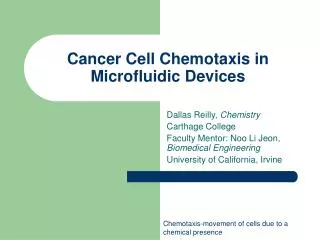
The Big Picture • Our microfluidic devices can be used to study chemotaxis, directed migration, of cell systems such as cancer • For example, it has been shown that breast cancer metastasizes based on epidermal growth factor (EGF) released from secondary target organs - we are able to study the details of this chemotaxis Chemotaxis: The characteristic movement or orientation of an organism or cell along a chemical concentration gradient either toward or away from the chemical stimulus Metastasis: A secondary cancerous growth formed by transmission of cancerous cells from a primary growth located elsewhere in the body
The Big Picture, cont. • The device I have been working with is easier to use than previous devices and is ideal for isolating individual cells • Isolating cells assists in studies that might target metastatic versus non-metastatic cells for gene research
Microfluidic Chemotaxis Chambers • Soft Lithography (a) A small patterning polydimethylsiloxane (PDMS) piece with embossed surface pattern is placed on a substrate that is coated with a thin film. (b) Exposure to reactive oxygen plasma selectively removes material in regions where the patterning piece does not contact the substrate. (c) After the patterning PDMS piece is removed, well-defined surface micropatterns of cell-adhesive or non-adhesive materials that can be used for selective cell attachment and growth. (d) A microfluidic PDMS piece with microchannel is aligned and bonded to the patterned substrate. The finished device can be used to culture patterned cells inside a microfluidic device. Seog Woo Rhee, Anne M. Taylor, Christina H. Tu, David H. Cribbs, Carl W. Cotmanand Noo Li Jeon. Patterned cell culture inside microfluidic devices. Lab on a Chip, 2004, 4.
Microscopes Inverted Differential Interference Contrast (DIC) Hoffman Modulation Contrast
Using the device • Making gradients -Use fluorescent beads and fluorescein isothiocyanate-dextran (FITC-Dextran) to image flow -Altered device assembly, timing of solution input, and flow rates to create gradients
Making Adjustments Beads Beads FITC-D FITC-D outlet (withdraw) outlet (withdraw)
Cell culture Growth of cells in vitro on an artificial medium for experimental research Cell Passaging The process of passing or maintaining a group of microorganisms or cells through a series of hosts or cultures Dilutions, confluence Experimentation Trypsinization of adherent cells Cell counting Cell starvation Addition of epidermal growth factor (EGF) Adding cells
Adding cells, cont. • Modeled protocol of paper our lab group published in 2004 using MDA-MB-231 metastatic breast cancer cells (~15-20 µm) Shurgen Wang, Wajeeh Saadi, Francis Lin, Connie Minh-Canh Nguyen, Noo Li Jeon. Differential effects of EGF gradient profiles on MDA-MB-231 breast cancer cell chemotaxis. Experimental Cell Research. 2004. • Slow movement of cells requires long experiment times – constant flow • Found that the high speeds needed to create gradient used up reservoir too quickly
Expanding reservoirs • Punch larger inlets • Thicker devices • Extend upwards - bonding reservoir walls • Tubing to attach pipette tips
Collaboration *Bonus points • Neutrophils -A type of white blood cell, specifically a form of granulocyte, filled with neutrally-staining granules, tiny sacs of enzymes that help the cell to kill and digest microorganisms it has engulfed by phagocytosis. *Movie
Smaller (~10-15 µm) Fast moving - Shorter experiment run times Short lived - Lots of tests in one day Use devices with smaller channels How to add more solution during test Make LOTS of devices Neutrophils
New Cell Lines • MTLn3 CFP (rat mammary adenocarcinoma cells with cyan fluorescent protein markers) -Background research -Changes in culturing -Impact of confluence -Alter experiment protocol
Changes in Protocol • Coating devices (cell attachment) - Fibronectin, Collagen I, Collagen IV - concentrations - Bovine Serum Albumin (BSA) • Cell starvation (induce movement) • Inside or outside device • How long • Channel size • Inducing chemotaxis - Growth factors, chemokines, extracellular matrix components - Concentrations • Cell speed • Experiment running time
Optimization Experiments • Cell adhesion • Cell movement • Cell shape • Cell velocity
Optimization Experiments • Diffusion versus suction for coating and cells • Getting cells into the channels • Number of cells
Altering the Device Buffer Buffer EGF + FITC-D EGF + FITC-D outlet (withdraw) outlet (withdraw)
Where to now • Now that set up appears to be consistent and cells are moving -Find optimal ranges of: EGF concentration; collagen concentration; cell number
Design devices with which to study these systems Make devices user friendly and design consistent protocols so data can be replicated Technology later applied to other fields Documentation of the optimal gradients, chemotactic characteristics and limiting factors in these systems will aid the development of treatment options to limit the directing effects of identified soluble factors Importance of this research
Acknowledgements • Mentor: Dr. Noo Li Jeon • Lab: Delaram Sahebzamani, Bobak Mosadegh, Madelyn Luttgen, Wajeeh Saadi, Bonggeun Chung, Jeong Won Park, Cyrus Roushan • IM SURE: Said Shokair, Jerry McMillan, Goran Matijasevic • National Science Foundation (NSF)