Download
1 / 18
180 likes | 737 Views
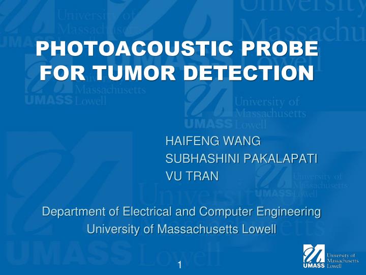
PHOTOACOUSTIC PROBE FOR TUMOR DETECTION. HAIFENG WANG SUBHASHINI PAKALAPATI VU TRAN Department of Electrical and Computer Engineering University of Massachusetts Lowell. OUTLINE. Introduction Principle of Ultrasound What is Photoacoustic (PA) Fiber-optic Source and Receiver

E N D
PHOTOACOUSTIC PROBE FOR TUMOR DETECTION HAIFENG WANG SUBHASHINI PAKALAPATI VU TRAN Department of Electrical and Computer Engineering University of Massachusetts Lowell
OUTLINE • Introduction • Principle of Ultrasound • What is Photoacoustic (PA) • Fiber-optic Source and Receiver • System of PA • How to fabricate • How PA system can detect tumor • Advantages and Disadvantages • Timeline • Tasks • Reference
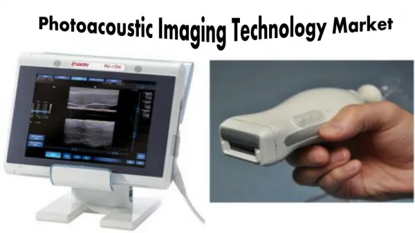
INTRODUCTION • Cancer killed ~570 000 people in 2010 in the USA. • Lot of methods to detect/predict tumor: US, x-ray, CT, MRI, PET, SPECT, etc. • Photoacoustic (PA) takes advantage of fiber optic and ultrasound to detect tumor
PRINCIPLE OF ULTRASOUND • Transducer emit ultrasound wave and get signals back from object • D= t.v • Scan volume to get image
WHAT IS PHOTOACOUSTIC? • Conversion of photons to acoustic wave due to absorption and localized thermal excitation. • Pulses of light is absorbed, energy will be radiated as heat. • Heat causes detectable sound waves due to pressure variation.
DIAGRAM OF A TYPICAL PA SYSTEM • Themo-elastic Ultrsound Generation • Extrinsic fiber hydrophone, based on a Fabry-Perot interferometer
FIBER OPTIC ULTRASONIC SOURCE BASED ON OPTO-ACOUSTIC CONVERSION
FIBER OPTIC ULTRASONIC RECEIVER BASED ON ACOUSTO-OPTIC CONVERSION
TRANSMISSION STRUCTURE 2D gold nanoparticle array sandwiched between a transparent substrate and a PDMS layer
GOLD NANOSTRUCTURE The top view (a) and sketch of the side view( b) of the gold nanostructure.
DETECTION OF TUMOR USING PAI • Tumors have dense and unorganized vasculature. • High density of blood vessels in tumors enhances PAI contrast.
DETECTION OF TUMOR (CONT.) Endogenous PA contrast: • Melanin • Oxy Hemoglobin • Deoxy Hemoglobin Exogenous PA contrast (Highly sensitive to deeply situated tumors): • Au NP • Alexa Fluor • Indocyanine Green
ADVANTAGES DISADVANTAGES • Ability to detect deeply situated tumor and its vasculature • Monitors angiogenesis • High resolution • Compatible to Ultra Sound • High Penetration depth 1. Limited Path length 2. Temperature Dependence 3. Weak absorption at short wavelengths
TASKS • HAIFENG WANG • Principle of Photoacoustic • Fabricate PA structure • SUBHASHINI PAKALAPATI • Principle and procedure of tumor detection using PA • AuNP enhanced • VU TRAN • Compare other methods to detect tumor • AuNP enhanced
REFERENCES [1] Jemal,A. et al. Cancer Statistics, 2010. CA CancerJ.Clin. 60, 277–300 [2] Hou, Y., et al., Optical generation of high frequency ultrasound using two-dimensional gold nanostructure. Applied Physics Letters, 2006. 89(9): p. 093901-3. [3] Mallidi, S., G.P. Luke, and S. Emelianov, Photoacoustic imaging in cancer detection, diagnosis, and treatment guidance. Trends Biotechnol, 2011. [4] Tang, M.X., et al., Photoacoustics, thermoacoustics, and acousto-optics for biomedical imaging. Proc Inst Mech Eng H, 2010. 224(2): p. 291-306. [5] Wang, X., et al., Photoacoustic tomography: a potential new tool for prostate cancer. Biomed Opt Express, 2011. 1(4): p. 1117-1126.