Download
1 / 21
290 likes | 992 Views
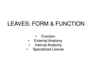
Leaves: Classification, Structure and Function. Functions a.) Photosynthesis - l eaves are the main photosynthetic organs of most plants (some green stems are also photosynthetic.) - flat, wide leaves have a lot of SA to trap sunlight. - upper surface of leaf is chlorophyll rich.

E N D
Leaves: Classification, Structure and Function
Functions a.) Photosynthesis - leaves are the main photosynthetic organs of most plants (some green stems are also photosynthetic.) - flat, wide leaves have a lot of SA to trap sunlight. - upper surface of leaf is chlorophyll rich
b.) Storage – some food and water is stored in the leaves ex. Lettuce leaves store water c.) Reproduction – asexual ex. cuttings d.) Transportation – of sugars to the stems - of water via transpiration
● Classification of Leaves While leaves vary extensively in form, they generally consist of a flattened blade and a stalk, the petiole, which joins the leaf to a stem node. In the absence of petioles in grasses and many other monocots, the base of the leaf forms a sheath that envelops the stem. Plant taxonomists use leaf shape, spatial arrangement of leaves, and the pattern of veins to help identify and classify plants.
a.) Venation Most monocots have parallel veins that run the length of the blade without joining one another, while dicot leaves have a multi-branched network of major veins or net venation.
b.) Types of Leaves • For example, simple leaves have a single, undivided blade, while compound leaves have several leaflets attached to the petiole. Fig. 35.5
Some plants have leaves that have become adapted by evolution for other functions. • This includes tendrils to cling to supports, spines of cacti for defense, leaves modified for water storage, and brightly colored leaves that attract pollinators. Fig. 35.6
Structure of Leaf Upper epidermis • Palisade Mesophyll– primary site for photosynthesis, just under upper epidermis. - contain many chloroplasts - cells are shaped like bricks and tightly packed • Spongy Mesophyll – promotes rapid diffusion of gases because of large air spaces - irregularly shaped and randomly arranged - fewer chloroplasts C. Stomata
Overhead view of stomata • C. Stomata • – are tiny pores in the epidermis layer of the leaf that regulate • gas exchange (CO2 in and O2 out) • allow for transpiration (loss of water vapour to occur) • the stomata are bordered by a pair of suasage shaped guard • cells that regulate the opening and closing of the stomata • when water levels are low, the guard cells become limp & rest • against one another, thus closing the stomata • when water levels are high, cells swell, thus opening the stomata
Typical Daily Guard Cell Schedule • Morning • Stomata open as photosynthesis begins in the chloroplasts • Oxygen level in leaves is greater than in the air • Gas exchange occurs by diffusion & water vapour is lost • Afternoon • Water concentration continually drops & cells shrink • Cells become limp & close • Night • Water builds up & stomata open • Stomata opening & closing is also related to: • CO2 concentration in guard cells, light levels, temperature & ABA • (abscisic acid concentration)
d.) Veins • Contain the vascular tissue – xylem and phloem (called vascular bundles)
Monocot Leaf Cross-Section • Given the number of vascular bundles in monocot stems, it should not be a surprise to see the numerous veins in their leaves. • Each vascular bundle represents 1 vein. • The mesophyll tissue most often isn’t differentiated into two distinct layers like in the dicot leaf.
Dicot Leaf Cross-Section • Dicot Leaves usually have a large central midrib which contains a large midvein (vascular Bundle). Minor Veins branch from the midvein at oblique angles • Dicot Leaves typically have two kinds of parenchyma (Palisade & Spongy) clearly present.
Cross Section of Dicot Leaf
Cuticle Upper Epidermis Palisade mesophyll Vascular Bundle Spongy mesophyll Lower Epidermis