Download
1 / 48
490 likes | 500 Views
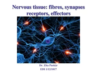
Nervous tissue : fibres , synapses receptors , effectors. Dr. Zita Puskár EDI 30/11/1018. Axons. Myelinated (enclosed within special sheet). Unmyelinated. Myelinated axon. Myelin sheath in the CNS. The myelin sheat is formed by the processes of oligodendrocyte.

E N D
Nervoustissue: fibres, synapsesreceptors, effectors Dr. ZitaPuskár EDI 30/11/1018
Axons Myelinated (enclosed within special sheet) Unmyelinated
Myelin sheath in the CNS The myelin sheat is formed by the processes of oligodendrocyte. One oligodendrocyte myelinate parts of several axons.
Composition of myelin MBP: Myelin basic protein PLP: Proteolipid protein 40% water, 70-85% lipid és 15-30% protein
Myelination in the periphery internodium Axons are sheated by Schwann cells.
A portion along the axon sheated by the Schwann cell is the internodal segment. Unmyelinated part of the axon between two internodal segments is the node of Ranvier. The node of Ranvier is covered by basement membrane produced by the two neighbouring Schwann cells. Schmidt-Lantermann clefts: nexus, punctum adherens → diffúsion path, maintenance
Unmyelinated axons PNS CNS Peripheral Nervous System (PNS) More than one axon is engulfed by Schwann cells Central Nervous System(CNS) Free axons. Only astrocyte endfeets are attached to them. They are not sheated at all.
Nerve fibers Nerve fiber: Axon (neurit) + glial sheath (if exists) Boundles of nerve fibers form tracts in the CNS
Boundles of nerve fibers form the peripheral nerve in the periphery Fascicles Sensory and motor fibers are mixed generally in the peripheral nerve
Connective tissue sheaths of the peripheral nerve Endoneurium: loose connective tissue with reticular fibres around each fibre Perineurium: myofilament containing, flat, epithelial like cells among circularly arranged connective tissue fibres around each fascicles (1-10 layers). The cells attached to each other by tight junctions and form diffusion barrier. Blood vessels are located in this layer. Epineurium: connective tissue sheath containing long, wavy collagen fibres around the nerve. It contains larger blood vessels. Paraneurium*: loose connective tissue.
Peripheral nerve fibers Conduction velocity and diameter A fibers B fibers C fibers 1-20 µm Ø 1-3 µm Ø 0,5-1 µm Ø 70-120 m/s 3-60 m/s 0,5-2 m/s M S M S,N Pr N, Po M: motor, S: Sensory, Pr: preganglionic N: nociceptor, Po:postganglionic
Interneuronal communication: synapsis Classification: (1) pre-and postsynaptic elements :axo-axonic, axo-dendritic, axo-somatic, dendro-dendritic , (2) modalities of the transmission (electronical, chemical), (3) postsynaptic effect (excitatory, inhibitory), (4) morphology: simple, composed
Axo-dendritic, axo-somatic, axo-axonic, dendro-dendritic szinapses
Electrical and chemical synapses bidirectional
Electrical synapses Connexon-6 connexin Synaptic cleft: 2 nm Exchange of ions, small molecules Bidirectional connection Sensory cortex, cerebellar cortex, retina, bulbus olfactorius
Chemical syanpses Composition: presynaptic element (axon terminal) • vesicles containing neurotransmitter, • presynaptic membrane, synaptic cleft Postsynaptic element (soma, dendrite) Postsynaptic membrane specialization with receptors Synaptic cleft: 20 nm Unidirectional connection
Simple or composed synapses Simple glomerulus (composed) thalamus, spinal cord
Classification of sensation Special Special sense organ Vision, hearing, balance, sense of smell or taste General/Somatic No special sense organs touch, thermal, pain (nociception), movement mozgás Somatic sensation Exteroceptor: information from the outside world Proprioceptor: information about the skeleto-muscular system, deep tissues Interoceptor: information about the state of the internal organs Sherrington (1906)
Sensation - Perception - Response Central Nervous System somatosensory afferents viscerosensory afferents visceromotor efferents somatomotor efferents receptor effector stimulus skin, muscle tendon (outside world) viscera glands, smooth muscles striated muscles
Receptor molecules proteins small molecules light ions Receptors – special protein complexes Location: cell membrane, cytoplasm, nucleus Activation: binding of specific molekules (ligands) or direct energy (heat) Consequences: ion movement and/or activation of second messanger systems resulting in specific cell response. stimulus → receptor activation (transduction) → (nerve)impulse
Secondary sensory epithelium Receptor cell Primary sensory epithelium Olfactory epithelium Hair cells Taste bud
Free nerve ending Thin myelinated (Aδ) or unmyelinated (C)afferent nerve endings containing complex receptor molecules. It can be thermo-, cold receptor or nociceptor (responds to damaging or potentially damaging stimuli)
Nociceptors Polimodal nociceptor Stimuli: mechanical, heat, chemical Mechano-cold nociceptor Stimuli: mechanical, cold Insensitive to mechanical stimuli Michael S. Gold & Gerald F Gebhart (2010) Nature Medicine, 16: 1248-1257
Mechanoreceptors of the skin Merkel Meissner Vater-Pacini Ruffini
Tactile (Merkel) Cell Mechanoreceptorcellsthatareactivatedby (light) pressure. Theyarelocatedinthestr. basaleof theepitheliumcontaining keratin and fewmelanosome. Basolateralsurface of thecellscontacttheterminaldiscsofsensoryfibers(Aβ).
Tactile (Meissner) - Corpuscle Mechanoreceptorthat is activatedbylightpressure (touch). It is an ellipsoidshapestructureenclosedby a connectivetissuecapsule (40x100 μm). MC is locatedinthedermalpapillae. Itconsists of 5-10 of flattened layers of cells, each with an elongated nucleus. The (Aβ) nerve endingloosingitsmyelinsheathis coiled among these cells.
Ruffini Corpuscle Ruffini’s present to Sherringtonnak Oxford University It is encapsulated cilindrical structure. The RC is a tension (mechano) receptor. The structure is very similar to the Golgi tendon.
Vater-Pacini corpuscle It is an encapsulated mechanoreceptor that is activated by course or sustained touch and vibration. It consists of connective tissue capsule with elastic fibres, outer lamellar and inner cylindrical structure. Lamellae: perineural cells (50-60 layers) and gel. Inner cylinder: unmyelinated nerve ending surrounded by circularly arranged Schwann cell layers and fluid. Location: dermis, joints, tendons
Krause - corpuscle Encapsulated mechanoreceptor. It is activated by tension. (Location: lips, tongue and genitals (erogen zone).
Muscle spindle (stretch receptor) Detects changes in the length of the muscle → regulation of the contraction of the muscle (stretch reflex)
Effectors Smooth muscle Striated muscle Glands
Irodalom: Röhlich Pál: Szövettan. Budapest, 2006 Eric R. Kandel, James H Schwartz and Thomas M Jessel: Principle of neural science, New York, 2000 Anthony L. Mescher: Junqueira’s Basic Histology, New York, 2010 Michael Ross and Lynn J. Romrell: Histology, Baltimore, 1989