Download
1 / 18
240 likes | 804 Views
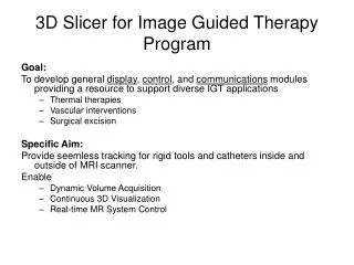
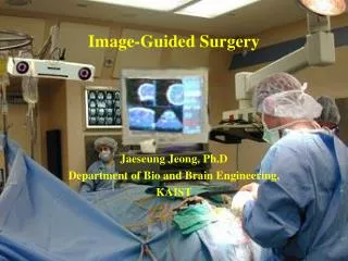
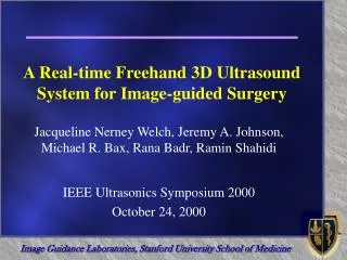
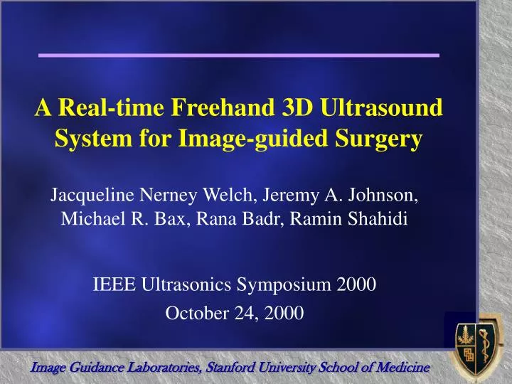
A Real-time Freehand 3D Ultrasound System for Image-guided Surgery. Jacqueline Nerney Welch, Jeremy A. Johnson, Michael R. Bax, Rana Badr, Ramin Shahidi. IEEE Ultrasonics Symposium 2000 October 24, 2000. Overview. Design motivations and decisions 3D ultrasound Freehand scanning

E N D
A Real-time Freehand 3D Ultrasound System for Image-guided Surgery Jacqueline Nerney Welch, Jeremy A. Johnson, Michael R. Bax, Rana Badr, Ramin Shahidi IEEE Ultrasonics Symposium 2000 October 24, 2000
Overview • Design motivations and decisions • 3D ultrasound • Freehand scanning • Optical tracking • Volume rendering • Simultaneous acquisition and visualization • Methods • Equipment • Spatial calibration • Volume construction and maintenance • Results • Future Work
Ultrasound • Ultrasound versus other imaging modalities (CT, MR, X-ray) • Least expensive • No ionizing radiation • Compatible with existing surgical instruments • Widely available and commonly used • Real-time, interactive nature
3D Visualization of Ultrasound • Compared to 2D, 3D provides: • More intuitive and comprehensible images • More accurate volume estimation • Shorter scanning times • Improved sharing of information 2D Ultrasound Image Volume Rendered 3D US
k (x,y,z) i j 3D from Conventional 2D Ultrasound 2D Images Volume Construction Engine Position Data Volume Rendering Engine Workstation US Probe Tracking Device
Optically Tracked Freehand Acquisition • Freehand versus other scanning techniques (mechanical) • Greatest freedom of movement • Compact • Least cumbersome • Requires probe position measurements • Optical versus other position tracking methods (magnetic, mechanical, speckle decorrelation) • Insensitive to metallic surgical equipment • Allows volume localization
Interactive Volume Rendering • Volume rendering versus other visualization methods (slice projection, surface rendering) • Truest to the data set • Easiest to interpret • Segmentation not required • Computationally expensive but feasible with current technology
Static Volume Volume Construction Engine k k (x,y,z) (x,y,z) Visualization Data Storage i i j j Dynamic Volume Volume Construction Engine Simultaneous Acquisition & Visualization Simultaneous Acquisition & Visualization Acquisition
Equipment • Image Guided Technology FlashPoint™ 5000 optical tracking system with 580 mm camera • Sonosite handheld ultrasound scanner with 5MHz linear probe • SGI 320 Visual Workstation with a single processor running Windows NT
Calibration Parameters kP • 6 extrinsic parameters • Rotation (Ri , Rj , Rk) • Translation (ti , tj , tk) • 2 intrinsic parameters • Image scale (si , sj) • Can be written as Probe Tracking Device Coordinates iP jP (Ri , Rj , Rk) (ti , tj , tk) (si , sj) iS, u jS, v Slice Coordinates
Calibration Phantom Image of Phantom During Calibration Ultrasound Phantom (1/16” Acrylic)
Calibration Method • Obtain feature positions • Align ultrasound probe • Capture US image and probe position • Localize features in image • Calculate calibration parameters • Scale factor • Rotation and Translation
Volume Construction and Maintenance Insertion of New Slices Removal of Old Slices Overwrite Existing Slices Interpolate with Nearby Slices
Future Work • Quantify and improve system performance • Spatial and temporal accuracy • Data rates • Display position and trajectory of surgical instruments • Apply system to clinical situations
Acknowledgements • Dr. Thomas Krummel’s lab • DOD Graduate Research Fellowship • CBYON, Inc.