Download
1 / 11
220 likes | 2.72k Views
Cor triatriatum dexter. James Montgomery, DVM October 19, 2009. Chester Acc #76718. 2 yo M Lab retriever Heart murmur, lethargic. Cor Triatriatum. Sinister and/or dexter Reported in both cats and dogs Dexter results from persistence of the embryologic right sinus venosus valve

E N D
Cortriatriatumdexter James Montgomery, DVM October 19, 2009
Chester Acc #76718 • 2 yo M Lab retriever • Heart murmur, lethargic
CorTriatriatum • Sinister and/or dexter • Reported in both cats and dogs • Dexter results from persistence of the embryologic right sinus venosus valve • The right atrium is partitioned by a diaphragm • Typically, the caudal vena cava and fossaovalis are in the caudal chamber and the coronary sinus and right AV valve are in the cranial chamber • Perforate or imperforate • Imperforate generally shunt to azygous or vertebral venous circulation
Cortriatriatumdexter • Extremely uncommon • Age at diagnosis range in lit from 8 weeks to 7 years • Reported breeds: chow chow, cocker spaniel, English bulldog, German shepherd dog cross, German short-haired pointer, golden retriever, greyhound, rottweiler, and mixed breed dog • Should be considered as a differential diagnosis in young dogs with abdominal distension and ascites
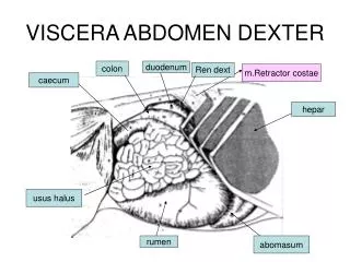
Clinical Findings • Usually have evidence of congestion in the caudal half of the body – ascites, but do not demonstrate jugular distension or a murmur • Prominent cutaneous abdominal veins • Hepatomegaly • Diarrhea from malabsorption
Diagnostics • Thoracic radiographs: tend to be unremarkable with the exception of caudal vena caval enlargement • ECG: generally normal • Diagnosis usually by echocardiography or selective angiography
Treatment • Balloon Valvuloplasty • Surgical resection or bypass of the obstruction
References • Duncan RB, et al. Cortriatriatumdexter in an English bulldog puppy: case report and literature review. J Vet Diagn Invest 11:361-5 (1999). • Oyama MA, et al. Congenital heart disease. In Ettinger SJ, Feldman EC, eds. Textbook of Veterinary Internal Medicine, 6thed (St. Louis, MO: Elsevier Saunders, 2005) pp. 1012-13.