Download
1 / 28
340 likes | 1.32k Views
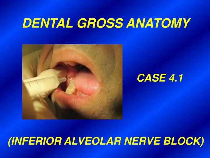
DENTAL GROSS ANATOMY CASE 4.1 (INFERIOR ALVEOLAR NERVE BLOCK). HISTORY A 23 yo man went to a dentist to have a mandibular 3 rd molar extracted. The patient requested that plenty of anesthetic be given because he was extremely sensitive to pain.

E N D
DENTAL GROSS ANATOMY CASE 4.1 (INFERIOR ALVEOLAR NERVE BLOCK)
HISTORY • A 23 yo man went to a dentist to have a mandibular 3rd molar extracted. • The patient requested that plenty of anesthetic be given because he was • extremely sensitive to pain. • The dentist inserted the needle through the mucous membrane on the • inside of the patient’s mouth, just lateral to the ridge produced by the • underlying pterygomandibular raphe. After penetrating the adjacent • muscle the tip of the needle came to rest near the lingula. • In a few minutes the patient stated that his gum, lower lip, chin and tongue • on the affected side were numb. • During the extraction the patient said he felt pain; the dentist injected more • anesthetic. The tooth was removed without further incident. • As the patient was leaving the dentist’s office he happened to look in a • mirror and was surprised to find that he was unable to close his eye and • that his mouth sagged on the affected side. He also noticed that his ear • lobe was numb. • The dentist explained that because of the large amount of anesthetic • injected, other nerves in addition to those supplying the teeth had been • anesthetized. He assured his patient that these effects would disappear • in 3-4 hours.
Name the nerve supplying the mandibular • 3rd molar tooth. What is this nerve a branch • of (be specific)? In what region of the head • does it arise?
NERVE SUPPLY OF THE TEETH V3 (POST. DIVISION) INFERIOR ALVEOLAR N.
The pterygomandibular raphe is used as a • landmark when giving this type of nerve • block. What is the pterygomandibular • raphe and what are its bony attachments?
Pterygoid hamulus Buccinator m. Pterygomandibular raphe Superior pharyngeal constrictor m. Mandible
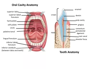
PTERYGOMANDIBULAR FOLD CAN BE SEEN AND PALPATED Maxillary anterior teeth Pterygomandibular fold Uvula Dorsum of tongue
What muscle was penetrated by the • needle?
What is the lingula of the mandible? • What fibrous structure attaches to it? • What function does the lingula and the • fibrous structure have when giving this • type of nerve block?
Lingula Mandibular foramen
Why was the patient’s chin and • lower lip on the injected side also • anesthetized?
Temporalis fascia and m. Anterior division (V3) (mostly motor) Posterior and anterior deep temporal nn. Posterior division (V3) (mostly sensory) Foramen ovale Masseteric n. Lateral pterygoid n. and m. Auriculotemporal n. Chorda tympani n. Buccal n. Lingual n. Inferior alveolar n. (cut) Mylohyoid n. Mylohyoid m. (cut) Mental n. Inferior alveolar n. (cut) Digastric m. (anterior belly)
Why was the patient’s tongue on the • injected side numb? Specifically, what • part was affected? Were the tongue • muscles paralyzed? Why or why not?
Temporalis fascia and m. Anterior division (V3) (mostly motor) Posterior and anterior deep temporal nn. Posterior division (V3) (mostly sensory) Foramen ovale Masseteric n. Lateral pterygoid n. and m. Auriculotemporal n. Chorda tympani n. Buccal n. Lingual n. Inferior alveolar n. (cut) Mylohyoid n. Mylohyoid m. (cut) Mental n. Inferior alveolar n. (cut) Digastric m. (anterior belly)
INNERVATION OF TONGUE MUSCLES XII Styloglossus m. Genioglossus m. Hyoglossus m.
What is the relationship between the • sensory nerve identified above (#6) and • the mandibular 3rd molar? Is this nerve • liable to be injured by the clumsy • extraction of this tooth?
Lingual n. 3rd Molar
Describe the innervation of the gingiva • of the mandibular teeth. In view of your • answer, should the dentist have • anesthetized any other nerve before • performing this extraction?
What caused the patient’s facial paralysis • and loss of sensation in his ear lobule?
INFERIOR ALVEOLAR NERVE BLOCK Medial pterygoid m. Parotid gl. & facial n. Sphenomandibular ligament Lingual n. Inferior alveolar n. Pterygomandibular raphe Ramus of mandible Temporalis m. insertion Masseter m. Buccinator m.
What complication might have arisen • if the dentist had injected too far laterally? • Too far medially? (Hint: what muscle • might be pierced?)
INFERIOR ALVEOLAR NERVE BLOCK Medial pterygoid m. Parotid gl. & facial n. Sphenomandibular ligament Lingual n. Inferior alveolar n. Pterygomandibular raphe Ramus of mandible Temporalis m. insertion Masseter m. Buccinator m.