Download
1 / 15
180 likes | 365 Views
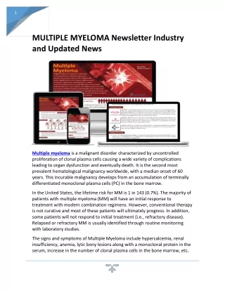
Multiple Myeloma. Morning Report July 21, 2009 Lindsay Kruska. Multiple Myeloma. Neoplastic proliferation of single clone of plasma cells producing monclonal immunoglobulin Cause unknown Radiation and solvents ?associated 1% malignancy, 10% hematologic malignancy in US

E N D
Multiple Myeloma Morning Report July 21, 2009 Lindsay Kruska
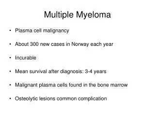
Multiple Myeloma • Neoplastic proliferation of single clone of plasma cells producing monclonal immunoglobulin • Cause unknown • Radiation and solvents ?associated • 1% malignancy, 10% hematologic malignancy in US • Incidence 4-5/100,000 • Median age presentation 60-66y, rare <40y (2%)
Presentation: Symptoms • Suspected usually due to widely varied symptoms (>6mo in 40%) • Bone pain (67%) • Weakness and Fatigue (30%) • Weight loss (24%) • Asthenia (14%) • Dyspnea (4%) • Fever (<1%) • Sx of cord compression • Repeated infections (pneumonia, pyelonephritis)
Presentation: Signs • Physical Exam • Pallor • Organomegaly, palpable LN rare (<5%) • Radiculopathy (cord compression) (5%) • Peripheral neuropathy uncommon • Labs • Hypercalcemia (36%) • Increased serum total protein • Anemia (34%) • Acute renal failure (34%) • Low anion gap
Work up • CBC with diff, smear • Anemia, rouleaux • Chemistries • Ca, Creatinine, Total protein • SPEP with immunofixation (87% sensitive) • UPEP (24h urine) (75% sensitive) • Serum light chains • Beta-2 microglobulin • Bone marrow biopsy • Bone survey, occasionally advanced imaging
SPEP Electrophoresis Immunofixation
Bone Marrow Biopsy • Marrow plasmacytosis (>10%) • CD138+, monoclonal • Focal BM involvement • 10% require multiple biopsies
Diagnosis • Presence of serum and/or urine monoclonal protein • IgG (53%), IgA (25%), IgD (1%) • Free light chains (20%) • Clonal plasma cells or plasmacytoma • End organ damage • HyperCa, Renal failure, Anemia, Lytic Bone lesions
Main Differential Diagosis: Elevated M protein • MGUS (1%/year) • Absence of symptoms • M protein <3g/L • <10% plasma cells in marrow • No anemia, renal failure, hyperCa, lytic lesions • Smoldering Multiple Myeloma (10%/year) • Meets dx criteria for MM but no end organ • Primary amyloidosis • Metastatic cancer
Other lymphoid neoplasms CLL B and T cell lymphomas Non lymphoid neoplasms Breast Ca CML Breast and colon cancer Cirrhosis Sarcoidosis Gaucher’s disease Pyoderma gangrenosum Autoimmune conditions Myasthenia gravis Rheumatoid arthritis Cold agglutinin disease Several rare skin DO Differential Also Includes
Staging • Durie-Salmon staging (Stages I-III, a/b), 1975 • M protein • Serum Calcium • Radiographic Bone Involvement • Hemoglobin • Renal failure • International Staging System (Stages I-III), 2005 • Serum beta2 microglobulin • Albumin
Therapy • Supportive care • Hypercalcemia/bone involvement: usual therapies, analgesia, occ XRT • Renal involvement: adequate hydration, ?plasmapheresis • Low threshold for infectious complications • Hyperviscosity: plasmapheresis • Neurologic compromise: palliative radiation • Anemia: transfusion • Initation of specific therapy • Chemotherapy • Thalidomide • Dexamethasone • Melphalan/prednisone • Bortezomib (proteasome inhibitor) • HSCT
Prognosis • Usually fatal • 10-20% mortality within first 2 months • Mean survival 4-5y • 5y survival 31%, 10y 10%, 15y 4% • Delay in diagnosis associated with negative impact on disease course • Lead time bias • Improved survival with therapy including HSCT • Major causes of death: progressive myeloma, renal failure, sepsis, therapy related acute leukemia or myelodisplasia; 25% die of age related illnesses
References • Fauci, et al. Harrison’s Internal Medicine. 17th edition. • Abeloff, et al. Abeloff's Clinical Oncology, 4th ed. • Up to Date. www.utdol.com