Download
1 / 85

E N D
1. Cerebellopontine Angle Tumors: Diagnosis and Management
Andrew Coughlin, MD
Tomoko Makishima, MD
University of Texas Medical Branch
Department of Otolaryngology
Grand Rounds Presentation
September 30, 2010
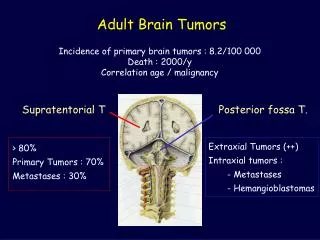
2. Outline History of CPA tumors
Discuss Relevant Anatomy
Epidemiology and Tumor Biology
Signs/Symptoms of CPA tumors
Brief overview of other CPA tumors
Treatment options
Present a case presentation
3. Overview of CPA Tumors Represent 10% of all intracranial tumors
Vestibular Schwannomas/Acoustic Neuroma represent 78% of these tumors (1)
Differential
Meningiomas
Epidermoids
Other Cranial Nerve Schwannomas
Dermoid Tumors ( Chordomas, Teratomas)
Arachnoid Cysts
Lipomas
Metastatic Tumors
Vascular Tumors (Hemangioma/Glomus Tumor)
4. History of CPA Tumors Sir Charles Balance (2,3)
1894 First successful removal of Vestibular Schwannoma
1907 Patient still alive but had CN VII paresis
Harvey Cushing
1917 Monograph described CPA syndrome
Pioneered subtotal resection through bilateral suboccipital craniectomy
5. History Continued Walter Dandy (5)
Advocated debulking with capsule removal and decreased operative mortality to 10%
William House (6-8)
Advocated Translabyrinthine approach 1960�s
Introduced the microscope and dental drill to identify the facial nerve
Introduced middle cranial fossa approach
6. CPA Syndrome (4)
7. Anatomy
8. Epidemiology Autopsy series have shown incidence of 1.7 to 2.7% (9,10)
MRI of 10,000 patients seen for non-otologic reasons showed 7 positive cases or 0.07% (11)
Denmark reports of 1.3 per 100,000 population (12)
9. Acoustic Neuroma Misnomer They are not Neuromas
They rarely are found on the acoustic portion of the nerve
*Involvement of the acoustic portion of the nerve often leads to erosion of the cochlea.
10. Tumor Biology Arise from Schwann cells within the IAC (1)
Equal frequency between inferior and superior divisions of the vestibular nerve.
Arise from Scarpa Ganglion instead of Obersteiner-Redlich zone.
Scarpa�s ganglion has highest density of schwann cells
11. Genetics NF 2 Gene found at 22q12
Tumor suppressor gene preventing schwann cell proliferation
Sporadic Variety (95%) (13)
Hypothesized that there are two hits to the normal NF gene
Neurofibromatosis type II
Autosomal Dominant
Inherit one abnormal and one normal gene with one hit to the normal allele.
Blood tests available to screen family members.
12. Biochemical Effects Neuregulin
Expressed by schwann cells to control proliferation and survival of schwann cells
Chemokines
FGF, TGF-�, VEGF, PDGF (14-17)
Sex Hormones (18)
Previous studies showed increased growth in pregnancy
Recent studies have not shown growth modulation or receptors for sex hormones
13. Workup History and Physical Exam
Audiologic testing
Vestibular Testing
Imaging
14. Symptoms Intracanalicular tumors
Hearing Loss
Tinnitus
Vestibular dysfunction/Vertigo
CPA extension
Disequilibrium/Ataxia
Brainstem Extension
Midface Hypesthesia
Hydrocephalus (vision loss and headache)
Other Cranial Neuropathies
*SNHL>tinnitus>disequilibrium>facial hypesthesia (13)
15. Unilateral Hearing Loss Present in >85% of patients (19)
5% of patients with VS have no associated hearing loss (20)
Speech discrimination out of proportion to HL
Many notice difficulty on the telephone
16. Association with SSNHL Occurs in up to 20% of patients (13)
1% of patients with SSNHL have a vestibular schwannoma (21)
48% of patients with SSNHL will have some recovery of hearing (21)
Don�t rule out VS if they recover
17. Tinnitus 2nd most common presenting sign
Often precedes hearing loss
Can be present without hearing loss
Can be high pitch, hissing, or a roar
Can localize to the opposite ear
Unilateral tinnitus should be evaluated
18. Vestibular Complaints 36-50% of patients describe disequilibrium (1)
Vague, transient lightheadedness
Acute vertigo is presenting symptom in 27% of patients but is associated with smaller tumors.
19. Facial Hypesthesia Presenting symptom in 4% of patients
Larger tumors >2cm
Maxillary division 1st
Corneal reflex is the first to go
Facial weakness is rare
If present should assume a different type of tumor.
20. Ocular Complaints Rare
Loss of corneal reflex
Nystagmus (toward affected side)
Diplopia
Involvement of CN VI
Blurry vision
Papilledema and optic atrophy from compression
21. Signs Hitselberger Sign
Decreased sensation of EAC
Sensory VII more sensitive than Motor VII
Absent corneal reflex, nystagmus
Hypesthesia to pinprick and touch
Weakness of Temporalis/Masseter muscles
Other cranial neuropathy�s
Gait disturbances or difficulty with finger to nose testing
22. Audiologic Testing Downsloping high frequency SNHL 65% (22)
5% of patients show no hearing loss (23)
Rollover: Speech discrimination worse than expected and worse with increased sound intensities
Retrocochlear losses more common than cochlear
23. Hearing Classification Scales (24)
24. Acoustic Reflexes **Positive test has 85% sensitivity for identifying retrocochlear problem
Acoustic Reflex Threshold
Increased (compared with cochlear norms) or absent if retrocochlear process
25. Auditory Brainstem Evoked Response Sensitivity of 85 to 90% (25)
False Positive rate 10%
False Negative rate 18-30% for intracanalicular tumors
Number larger than in the past
Five waveforms are produced with the most common being a latency in wave V compared to the normal ear of >0.2msec.
Recommended as a screening test for those with low suspicion of vestibular schwannoma.
26. Vestibular Testing 70-90% of patients will show some abnormality (26)
50% of small tumors produce no abnormalities (27)
Caloric testing is commonly the only abnormality
Inferior vestibular nerve tumors may be missed
Superior nerve showed decrease in 98%
Inferior nerve shows decrease in only 60% (28)
Therefore not used as a screening test
27. CT Imaging 90% of vestibular schwannomas will enhance with contrast
Frequently misses tumors that are not intracanalicular and do not extend >5mm to the CPA.
63% accuracy at diagnosis (29)
28. MRI Gold Standard for Vestibular Schwannomas
Can identify tumors as small as 3mm (30)
Gadolinium is preferentially taken up by tumor
Better visualization of small tumors
Gadolinium enhanced studies
Hyperintense on T1 and T2 studies
Non-contrasted studies
Hypointense T1, Isointense on T2
Some recommend T2 fast spin-echo MRI as screening test but most will likely require at thin slice MRI if abnormalities are found (31)
29. MRI continued Vestibular Schwannoma Meningioma Centered on IAC
Globular appearance
Ice cream cone appearance in IAC
Bony erosion of IAC
Cystic degeneration or hemorrhage may be present Extend along petrous ridge
Sessile appearance
�Dural Tail� at periphery
Iso/Hypointense on T1
Hypo to Hyperintense on T2
30. Acoustic vs Meningioma
31. Differential Diagnosis
32. Meningiomas 3% of all CPA tumors (13)
Do not metastasize but do recur due to bony invasion
Are formed from cap cells around arachnoid villi near dural sinus� and where CN�s enter their foramina
Generally are not intracanalicular so must be larger to cause hearing concerns
33. Meningioma Signs and Symptoms (32)
Usually cause spontaneous nystagmus, facial hypesthesia, and gait ataxia
If inferior can cause hoarseness, dysphagia, tongue atrophy
If within the IAC can produce similar symptoms as Vestibular Schwannoma
Hearing tests will show retrocochlear process if large enough, and 25% will have normal ABR�s (33)
34. Meningioma Treatment
Surgical Excision
Must remove rim of normal dura and possibly bone
Poor hearing
Translabyrinthine approach
Good hearing
Suboccipital or Middle Fossa Approach
Middle Fossa is better for more superiorly based tumors.
35. Epidermoids Identical to cholesteatoma
Develop from epithelial rest cells
Slow growing
Commonly don�t present until 20-30�s
Arise adjacent to brainstem and infiltrate areas of least resistance
Can have irregular shape and infiltrate widely
36. Epidermoids Signs/Symptoms
Facial twitching is commonly described
Can become quite large before causing symptoms
Progressive facial paralysis more common than schwannomas
Audio/ABR/Speech discrimination consistent with other retrocochlear losses
Treatment is surgical excision
37. MRI Epidermoid (13)
38. MRI Arachnoid Cyst (13)
39. Facial Schwannoma Histologically identical to Vestibular Schwannomas
Characteristics
Rarely restricted to IAC
Commonly have skip lesions
Generally involve part of geniculate ganglion
40. Facial Schwannoma Signs/Symptoms
Unilateral hearing loss
Tinnitus
Vertigo
Aural fullness (if distal to geniculate)
Facial weakness is rare unless very large
Hearing tests show retrocochlear process but impedance testing can show ipsilateral absent acoustic reflex
41. Facial Schwannoma Treatment is observation
If growth or facial nerve dysfunction
Resection of nerve with cable grafting
Translabyrinthine approach most commonly used
Facial nerve decompression can be used if paresis is developing
42. Other CPA Tumors (1) Glomus Tumors
Jugular Foramen Syndrome (IX,X,XI), Surgical excision
Hemangiomas
Centered on geniculate, slow progressive facial weakness, surgical excision with facial nerve grafting
Arachnoid Cysts
Treatment is drainage
Cholesterol Granulomas
Bright on T1 and T2, Drained via infralabyrinthine approach
Embryonic tumors (Dermoids, teratomas, chordomas)
Excision with dysfunction
Primary Axial Tumors of CNS (glioma, hemangioblastoma, medulloblastoma)
Surgical excision +/- radiation therapy
43. Management Observation
Surgery
Stereotactic Radiosurgery
Radiation Therapy
**Generally tumors <3cm can be treated with stereotactic radiosurgery but microsurgery is the treatment of choice
44. Observation Growth rates vary from 0 to 2cm per year with an average of 2mm (34)
38.9-82% have some growth at =38 months (24)
14 to 24% of patients watched will go on to have some treatment (12)
Age should not be determining factor
Young patient with small tumor
Older patient with large tumor
Older patient with small tumor
Single MRI is not adequate
Repeat at 6 months and then again yearly
Longstanding hearing loss may represent a slow growing tumor (35)
45. Radiation Therapy
46. Stereotactic Radiosurgery First Introduced Leskell in 1969
Gamma Knife
Uses 201 ionizing beams of gamma rays from cobalt 60 source
One session
LINAC
Uses multiple beams from a linear accelerator
One session
Fractionated Radiotherapy
Newer therapy to eradicate cells in different cell cycle stages
Multiple sessions
Goal is to stop tumor growth, not shrink or remove tumor
47. Stereotactic Radiosurgery (24) Local control = not requiring salvage therapy
87 to 100% local control rates all three combined
23% of cases have transient increase tumor volume due to central necrosis
6mo to 5yr to disappear
Therefore some patients receive unnecessary salvage therapy
48. Gamma Knife Hasegawa et al. 2005 (36)
317 patients
Median follow up 7.8 years
10 yr local control >92%
Partial or complete radiographic response 62%
Progression free survival
96% if <15cm3 vs 57% if >15cm3 p<0.001
Better with no brainstem compression/4th ventricle obstruction p<0.001
Most tumor progression within 3 years
49. LINAC Freidman et al. 2006 (37)
390 patients
Median follow up 40 months
Median dose of 12.5 Gy
5- and 10-year local control 90%
Only 1% of patients required surgery for treatment failure
50. FSRT (24) Many different regimens studied
Dose 15-57.6 Gy/3-32fx
Median Follow ups of 48 months
5- year local control of >90%
**Relatively new idea so no long term outcomes available
51. Radiosurgery vs FSRT (38) Tumor Control
73.8 to 100% for Radiosurgery
91.4 to 100% for FSRT
Tumor Shrinkage
38 to 76.2% for Radiosurgery with single dose
34 to 76% for FSRT
Tumor Growth
0-26.2% for Radiosurgery
0-12.5% for FSRT
Hearing Preservation
47-71% for Radiosurgery
57-100%for FSRT
52. Hearing Preservation Defined as G-R class I/II
Early studies 16Gy = 46%
Later Studies 12-13Gy = 68-78% with no change in control (24)
Prasad et al. (39)
153 patients
Median follow up of 4.2 years
Useful hearing preservation rate of 58%
Size matters
<1cm3 = 75% hearing preservation
>1cm3 = 57% hearing preservation
53. Hearing Preservation GK vs FSRT
Andrews et al. 2001 (40)
109 patients
63 GK at 12Gy
46 FSRT at 50Gy/25fx
Follow up was 3 years
33% vs 81% hearing preservation (GK/FSRT)
98% vs 97% local control (GK/FSRT)
Thought is that lower dose in multiple fractions leads to decreased late normal tissue damage
54. Facial Neuropathy GK dose decrease from 16Gy to 12-13Gy decreased rates from 15% to 0% (41)
LINAC dose decrease of 16Gy to 12.5Gy showed drop in neuropathy from 4.4% to 0.7% (37)
RS vs FSRT ranges of 0-52% vs 0-4%(38)
55. Trigeminal Neuropathy GK dose decrease from 16Gy to 12-13Gy decreased rates from 16% to 4.4% (41)
LINAC dose decrease of 16Gy to 12.5Gy showed drop in neuropathy from 3.7% to 0.7% (37)
RS vs FSRT ranges of 2.4-29% vs 0-16% (38)
56. Other complications Hydrocepahlus
0-11% (24), with highest rates in FSRT
Tinnitus
0.2 to 5%
Ataxia
1.4 to 3.6%
Vertigo
1.4 to 1.7%
Peritumoral cyst formation
3.6%
Malignant transformation
0 to 0.3%
3 cases in all studies reviewed with time to presentation of 5, 7.5, and 18 yrs post therapy
Disequilibrium
33% likely due to weaker central compensation (42)
57. Surgery Translabyrinthine Approach
Middle Cranial Fossa Approach
Retrosigmoid-Suboccipital Approach
**All will require discussion/collaboration with Neurosurgery
58. Translabyrinthine Approach (43) Advantages Disadvantages Least cerebellar retraction
Wide exposure of posterior fossa
No size limit for resection
Facial nerve easily identified throughout
Ease of facial nerve repair if damaged/resected during removal
Low recurrence
Low headaches Residual hearing is sacrificed
Requires abdominal fat graft
59. Middle Cranial Fossa Approach (43) Advantages Disadvantages Best hearing preservation
<30db PTA
>70% speech discrimination
Good exposure of lateral IAC, CPA, and clivus
Drilling is extradural decreasing morbidity Limited to tumors <2cm in greatest dimension
Temporal lobe retraction
Must dissect around facial nerve due to its superior position
Limited posterior fossa exposure
60. Retrosigmoid/Suboccipital Approach (43) Advantages Disadvantages Can attack any size tumor
Hearing preservation possible
Wide exposure of brainstem and lower cranial nerves
Neurosurgeon familiarity
Consistent facial nerve identification Must be medially located with <2cm CPA extension
Lateral tumors risk injury to endolymphatic sac and vestibular labyrinth
Cerebellar retraction occulomotor abnormalities and postop disequilibrium
Increased air embolism risk (Sitting vs Park Bench position)
61. Microsurgery Control Rates Translabyrinthine
Total Resection 99.5 to 99.7% (44-45)
Near total resection (<25mm2 or 2mm thick)
Visible on MRI in 50% of patients with 3% risk of recurrence (46-47)
Middle Cranial Fossa
Total Resection 98% (48)
Retrosigmoid
Total resection 95% (49)
62. Hearing Preservation Serviceable hearing defined as class A/B or 1/2 (24)
Middle cranial fossa 51%
Retrosigmoid 31%
Meyer et al. 2006 (50)
162 consecutive patients via MCF approach
Class A/B hearing preserved in 41%
56/113 (50%) with WR >70% preoperatively maintained that level.
Tumor 0.2-1.0cm = 59%
Tumor 1.1-1.4cm = 39%
Tumor 1.5-2.5cm = 33%
63. Facial Nerve Paralysis Reporting function at 6-12 month point is gold standard
Preserved = Grade I/II (24)
Retrosigmoid 91%
Middle Cranial Fossa 88%
Translabyrinthine 77%
Delayed paralysis (>72hrs after surgery)
Described by Grant et al. 2002
Incidence 5% (51)
79% regain postoperative function by 1 yr
64. CSF Leak Pooled data from 19 studies 360/4297 (8%) rate (52)
MCF approach 6%
TL approach 8%
RS approach 11%
Occurs at two different time points
POD 2-3 (Early)
POD 10-14 (Late)
65. Headache Occurs roughly 10% of patients >3 months postop (52)
RS approach 21%
MCF approach 8%
TL approach 3%
Mechanisms
Bone dust contacting CSF/Meninges ? Aseptic Meningitis
Occipital nerve entrapment
Scarred neck muscles and dura
Migraine
66. Other Microsurgical Complications (53) Mortality
1% due to neurovascular injury
Meningitis
1-8%
Aseptic more common than bacterial (bone dust/inflammation)
S. aureus most common pathogen
Tinnitus
50% with preop tinnitus will have resolution postop
Balance abnormalities
Occurs in most patients but gone by 6-9 months
Seizure/Hydrocephalus/Stroke (24)
Rare and <2% of cases
67. Microsurgery vs Radiosurgery Myrseth et al 2005 (53)
Retrospective review 189 patients tumors =3cm
86 by microsurgery vs. 103 by GK
5.9 year mean follow up
Local control rates of 89.2% Surgery vs 94.2% GK
HB 1-2 in 79.8% Surgery vs 94.8% GK p=0.0026
Quality of life significantly lower in surgery group compared to gamma knife group
*MCF approach was not utilized
68. Microsurgery vs Radiosurgery Pollock et al. 2006 (54)
Prospective cohort of 82 patients unilateral VS <3cm
36 Surgery vs 46 GK
Average follow up of 42 months
Local control 96% Surgery vs 100% GK p= 0.50
HB 1/2 in 75% Surgery vs 96% GK p<0.01
Hearing Preserved 5% Surgery vs 63% GK p<0.001
Quality of life all statistically better for GK
Physical functioning
Bodily pain scores
Dizziness Handicap Inventory
69. Summary Vestibular schwannomas are the most common CPA tumor
Unilateral Tinnitus or SNHL must be evaluated
Tumors <3cm in size treated with radiosurgery
GK or LINAC have comparable results
FSRT may prove more beneficial but requires more than 1 day of treatment
For patients with no serviceable preoperative hearing, translabyrinthine approach is best choice
For Hearing Preservation options the middle cranial fossa approach appears to be slightly better than the retrosigmoid approach due to decreased rates of complications.
70. Case Report 55 y/o female with 2 year history of falls and disequilibrium
Falls are now progressive and daily
Denies true vertigo just imbalanced
Also has tinnitus, neck spasms, and migraines
She denies any hearing loss but family feels otherwise
71. Case Report PMHx:
HTN, Hypothyroidism
PSHx:
Partial hysterectomy and R thyroid lobectomy
FHx:
Noncontributory
SHx:
45pack year history of smoking, -EtOH
Medications:
HCTZ, metoprolol, sydol prn, soma, and ambien prn
Allergies:
Sulfa
ROS:
Otherwise negative. No vision or constitutional problems
72. Case Report General: NAD oriented x3
Head: NCAT
Eyes: No nystagmus, PERRLA, EOMI
ENT: TM�s intact, Tuning forks normal
Neck: No masses or lymphadenopathy
Neuro: CN II-XII intact, unsteady Romberg, tandem gait falling to the left, and left finger to nose ataxia
73. Case Report Audiogram
Normal hearing with mild high frequency SNHL at 4000Hz bilaterally
ENG
Decreased accuracy on horizontal smooth pursuit
54% weakness on left air caloric testing
MRI
1.5 to 2cm lesion closely associated with CN VII/VIII. It is intracanalicular with extension into CPA. No evidence of brainstem compression or ventricular obstruction
74. Image 1
75. Image 2
76. Image 3/4
77. Treatment Plan? Finger to nose and gait ataxia
Essentially normal hearing
ENG positive for left lateral canal weakness
2cm tumor, no hydrocephalus
78. Case Report Patient had previous MRI 2005 showing no tumor
Therefore it is decided that we perform suboccipital craniotomy for excision of tumor
79. Images of Surgery
80. Images of Surgery
81. Images of Surgery
82. Images of Surgery
83. Case Report Final Pathology: Meningioma
No complications at this point
Tuning forks 256hz and 512hz normal
HB I immediately and 72 hours postoperatively
84. References Baileys.
House WF. A history of acoustic tumor surgery: 1800�1900, early history. In: House WF, Luetje CM, eds. Acoustic tumors Vol. 1. Baltimore: University Park Press, 1979:3�8.
Rosenberg SI. Natural history of acoustic neuromas. Laryngoscope 2000;110:497�508.
Cushing H. Tumors of the nervus acusticus and the syndrome of the cerebellopontine angle (Reprint of the 1917 edition.) New York: Hafner, 1963.
Dandy WE. An operation for the total removal of cerebellopontine (acoustic) tumors. Surg Gynecol Obstet 1925;41:129�148.
House WF. Report of cases: monographtranstemporal bone microsurgical removal of acoustic neuromas. Arch Otolaryngol 1964;80:617�667.
Glasscock ME III, Steenerson RL. A history of acoustic tumor surgery: 1961�present. In: House WF, Luetje CM, eds. Acoustic tumors Vol. 1. Baltimore: University Park Press, 1979:33�41.
House WF. Exposure of the internal auditory canal and its contents through the middle cranial fossa. Laryngoscope 1961;71:1363�1385.
Hardy M, Crowe SJ. Early asymptomatic acoustic tumor: report of 6 cases. Arch Surg 1936;32:292�301.
Eckermeier LS, Sorenson GD, McGavran MH. Histopathology of 30 non-operated acoustic schwannomas. Arch Otorhinolaryngol 1979;222:1.
Anderson TD, Loevner LA, Bigelow DC, et al. Prevalence of unsuspected acoustic neuroma found by magnetic resonance imaging. Otolaryngol Head Neck Surg 2000;122:643�646.
Tos M, Stangerup S, Caye-Thomasen P, et al. What is the real incidence of vestibular schwannoma? Arch Otolarygol Head Neck Surg 2004;130:216�220.
Cummings
Murphy PR, Myal Y, Sato Y, et al. Elevated expression of basic fibroblast growth factor messenger ribonucleic acid in acoustic neuromas. Mol Endocrinol 1989;3:225�231.
Diensthuber M, Brandis A, Lenarz T, et al. Co-expression of transforming growth factor-�1 and glial cell line derived neurotrophic factor in vestibular schwannomas. Otol Neurotol 2004;25:359�365.
Cay�-Thomasen P, Baandrup L, Jacobsen GK, et al. Immunohistochemical demonstration of vascular endothelial growth factor in vestibular schwannomas correlates to tumor growth rate. Laryngoscope 2003;113:2129�2134.
Brieger J, Bedavanija A, Lehr H, et al. Expression of angiogenic growth factors in acoustic neuroma. Acta Otolaryngol 2003;123: 1040�1045.
Beatty CW, Scheithauer BW, Katzmann JA, et al. Acoustic schwannoma and pregnancy: a DNA flow cytometric, steroid hormone receptor, and proliferation marker study. Laryngoscope 1995;105:693�700.
Selesnick SH, Jackler RK, Pitts LW. The changing clinical presentation of acoustic tumors in the MRI era. Laryngoscope 1993;103:431�436.
Sataloff�RT,�Davies�B,�Myers�DL:�Acoustic neuromas presenting as sudden deafness.��Am J Otol��1985;�6:349.
Friedman RA, Kesser BW, Slattery WH, et al. Hearing preservation in patients with vestibular schwannomas with sudden sensorineural hearing loss. Otolaryngol Head Neck Surg 2001;125:544�551.
Johnson EW. Results of auditory tests in acoustic tumor patients. In: House WF, Luetje CM, eds. Acoustic tumors Vol. 1. Baltimore: University Park Press, 1979:209�224.
Beatty�CW,�Ebersold�MJ,�Harner�SG:�Residual and recurrent acoustic neuromas.��Laryngoscope��1987;�97:1168.
Backous DD, Pham HT: Guiding patients through the choices for treating vestibular schwannomas: Balancing options and ensuring informed consent. Otolaryngol Clin N Am 2007; 40: 521-540.
Wilson DF, Hodgson RS, Gustafson MF, et al. The sensitivity of auditory brainstem response testing in small acoustic neuromas. Laryngoscope 1992;102:961�964.
Jenkins HA. Long-term adaptive changes of the vestibulo-ocular reflex in patients following acoustic neuroma surgery. Laryngoscope 1985;95:1224�1234
Nedzelski JM, Schessel DA, Pfleiderer A, et al. Conservative management of acoustic neuromas. Otolaryngol Clin North Am 1992;25:691�705.
85. References Linthicum FH. Electronystagmography findings in patients with acoustic tumors. Semin Hear 1983;4:47�53.
Welling DB, Glasscock ME III, Woods CI, et al. Acoustic neuroma: a cost-effective approach. Otolaryngol Head Neck Surg 1990;103:364�370.
Press�GA,�Hesselink�JR:�MR imaging of cerebellopontine angle and internal auditory canal lesions at 1.5T.��AJR Am J Roentgenol��1988;�150:1371.
Shelton C, Harnsberger HR, Allen R, et al. Fast spin echo magnetic resonance imaging: clinical application in screening for acoustic neuroma. Otolaryngol Head Neck Surg 1996;114:71�76.
Granick MS, Martuza RL, Parker SW, et al. Cerebellopontine angle meningiomas: clinical manifestations and diagnosis. Ann Otol Rhinol Laryngol 1985;94:34�38.
Selters WA, Brackmann DE. Acoustic tumor detection with brain stem electric response audiometry. Arch Otolaryngol 1977;103:181�187.
House�WF:�Translabyrinthine approach. ��In:�House�WF,�Leutje�CM,�Doyle�KJ,�ed.�Acoustic Tumors, �San Diego:�Singular Publishing;�1997:171-176.
Charabi S, Thomsen J, Mantoni M, et al. Acoustic neuroma (vestibular schwannoma): growth and surgical and nonsurgical consequences of the wait-and-see policy. Otolaryngol Head Neck Surg 1995;113:5�14.
Hasegawa T.,�Fujutani S.,�Katsumata S.,�et al:� Stereotactic radiosurgery for vestibular schwannomas: analysis of 317 patients followed more than 5 years. �Neurosurgery�57.�257-265.2005
Friedman W.A.,�Bradshaw P.,�Myers A.,�et al:� Linear accelerator radiosurgery for vestibular schwannomas. �J Neurosurg�105.�657-661.2006.
Likterov I, Allbright RM, Selesnick SH: LINAC radiosurgery and radiotherapy treatment of acoustic neuroma. Otolaryngol Clin N Am 2007; 40: 541-570.
Prasad D.,�Steiner M.,�Steiner L.:� Gamma surgery for vestibular schwannoma. �J Neurosurg�92.�745-759.2000.
Andrews D.W.,�Suarez O.,�Goldman W.,�et al:� Stereotactic radiosurgery and fractionated stereotactic radiotherapy for the treatment of acoustic schwannomas: comparative observations of 125 patients treated at one institution. �Int J Radiat Oncol Biol Phys�50.�(5): 1265-1278.2001.
Kondziolka D.,�Lunsford D.,�McLaughlin ,�et al:� Long-term outcomes after radiosurgery for acoustic neuromas. �N Engl J Med�1998, 339:�1426-1433.
Roland PS, Eston D. Stereotactic radiosurgery for acoustic tumors. Otolaryngol Clin North Am 2002;35:343�355.
Bennett M, Haynes DS: Surgical approaches and complications in the removal of vestibular schwannomas. Otolaryngol Clin N Am 2007; 40: 589-609.
Shelton C. Unilateral acoustic tumors: how often do theyrecur after translabyrinthine removal? Laryngoscope 1995;105:958�966.
Thedinger BA, Glasscock ME III, Cueva RA, et al. Postoperative radiographic evaluation after acoustic neuroma and glomus jugulare tumor removal. Laryngoscope 1992;102:261�266.
Pace-Balzan A, Ley RH, Ramsden RT, et al. Growth characteristics of acoustic neuromas with particular reference to the fate of capsule fragments remaining after tumor removal: implications for patient management. In: Tos M, Thomsen J, eds. Proceedings of the first international conference on acoustic neuroma Amsterdam:Kugler. 1992:701�703.
Bloch D, Oghalai JS, Jackler RK, et al. The role of less than complete resection of acoustic neuroma. Otolaryngol Head Neck Surg 2004;130:104�112.
Friedman RA, Kesser B, Brackmann DE, et al. Long-term hearing preservation after middle fossa removal of vestibular schwannoma. Otolaryngol Head Neck Surg 2003;129:660�665. 99.
Ebersold MJ, Harner SG, Beatty CW, et al. Current results of the retrosigmoid approach to acoustic neurinoma. J Neurosurg 1992;76:901�909.
Meyer T.A.,�Canty P.A.,�Wilkinson E.P.,�et al:� Small acoustic neuromas: surgical outcomes versus observation or radiation. �Otol Neurotol�2006, 27(3): 380-392.
Grant GA, Rostomily RR, Kim K, et al. Delayed facial palsy after resection of vestibular schwannoma. J Neurosurg 2002;97:93�96.
Harsha W.J.,�Backous D.D.:� Counseling patients on surgical options for treating acoustic neuroma. �Otolaryngol Clin North Am�2005, 38(4): 643-652.
Myrseth E.,�Moller P.,�Pederson P.H.,�et al:� Vestibular schwannomas: clinical results of quality of life after microsurgery or gamma knife radiosurgery. �Neurosurgery�2005, 56(5): 927-935.
Pollock B.E.,�Discoll C.L.W.,�Foote R.L.,�et al:� Patient outcomes after vestibular schwannoma management: a prospective comparison of microsurgical resection and stereotactic radiosurgery. �Neurosurgery�2006, 59(1): 77-85.