Download
1 / 13
130 likes | 285 Views
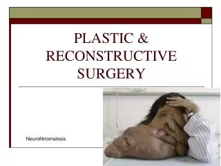
JAMA Facial Plastic Surgery Journal Club Slides: Free Tissue Transfer. Cannady SB, Rosenthal EL, Knott PD, Fritz M, Wax MK. Free tissue transfer for head and neck reconstruction: a contemporary review. JAMA Facial Plast Surg . Published online July 3, 2014. doi:10.1001/jamafacial.2014.323.

E N D
JAMA Facial Plastic SurgeryJournal Club Slides:Free Tissue Transfer Cannady SB, Rosenthal EL, Knott PD, Fritz M, Wax MK. Free tissue transfer for head and neck reconstruction: a contemporary review. JAMA Facial Plast Surg. Published online July 3, 2014. doi:10.1001/jamafacial.2014.323.
Introduction • Reconstruction in the head and neck follows a well-defined reconstructive ladder. • Microvascular free tissue transfer is usually reserved for problems at the apex of the ladder. • Numerous advances have been made both technically and from a tissue selection perspective. • Where free tissue transfer fits in the armamentarium of the facial plastic surgeon is a process in evolution.
Purpose • Objectives: • To provide a comprehensive review of the role of free tissue transfer in the management and rehabilitation of patients with maxillary and scalp defects. • To discuss the role of virtual surgical planning in head and neck reconstruction.
Relevance to Clinical Practice • Defects of the maxilla can be debilitating to the patient from cosmetic and functional perspectives. The ideal reconstruction will need to address osseous contour restoration, long-term soft-tissue volume restoration, separation of the oral and nasal cavities, and support of the eye. • Reconstruction of the bony contours of the mandible or maxilla can be difficult in the ablative setting. Virtual planning allows for determination of the best reconstructive plan in complex settings prior to the surgical extirpation. • Most lesions of the scalp can be closed primarily or with local tissue rearrangement. Occasionally one encounters a massive lesion or a lesion in a patient that has seen other treatments and local tissues are not available for reconstruction. In these patients, free tissue transfer may be the only option.
Description of Evidence • A comprehensive review of the literature was performed. Selected articles were reviewed and discussed. The authors’ personal extensive experience was used. • For maxillary reconstruction, there are many options available in the reconstructive paradigm. • If the infraorbital rim and orbital contents are left intact, then obturation should be considered. • While muscle flaps provide soft-tissue bulk, this bulk disappears over time. When combined with lack of a bony platform, the long-term results do not stand up. • The fibula flap is an excellent option for bony reconstruction as well as soft-tissue reconstruction. It also allows for dental rehabilitation.
Description of Evidence This patient has had maxillectomy that was reconstructed with a fibula free flap. Implants were placed later in the reconstruction.
Description of Evidence • Skin cancer is one of the most common forms of cancer in the head and neck. It is usually easily reconstructed with local flaps. • Patients with previous or anticipated radiation, very tight scalps, previous attempts at reconstruction with local flaps, or calvarial defects (full or partial thickness) may benefit from free tissue transfer in their reconstructive paradigm. • The latissimus dorsi myogenous flap with a split-thickness skin graft is the most commonly used flap in this scenario. • Cranial reconstruction can occur contemporaneously with the soft-tissue reconstruction.
Description of Evidence This patient has had a large scalp resection that was reconstructed with a latissimus dorsi myogenous free flap with a split-thickness skin graft. It has healed well and has an acceptable cosmetic outcome.
Description of Evidence • Mandibular reconstruction with composite bony flaps has been accepted as the reconstructive method of choice. • Preoperative virtual surgical planning is a technique that uses 3-dimensional computed tomographic scans with an interface over the Internet to plan the bony ablation and the bony reconstruction. • The planning allows for determination of the osteotomies and the manufacturing of surgical guides. • This in turn leads to a decrease in surgical time and better approximation of the segments. • In cases where a plate cannot be bent to the native mandible, the preoperative planning allows for the generation of a computer-designed reconstruction that facilitates oral rehabilitation.
Description of Evidence This patient will have a mandibular resection. The scans demonstrate the creation of the defect and how the bone will fit in the defect. The cutting jigs are also demonstrated.
Controversies and Consensus • Free tissue transfer is accepted as the best method of reconstructing composite tissue defects. • While obturators can be used in some maxillectomy defects, the use of free flaps allows for bony reconstruction. • Virtual modeling has improved the ability to preoperatively plan the reconstruction. However, the benefits on a cost basis are unknown. Also, it is unknown how this translates into functional benefits.
Comment/Conclusions • The role of free tissue transfer in head and neck reconstruction is well defined. Refinements in the use of this composite tissue to replace defects are an ongoing process. • The need to continue to improve the rehabilitation of patients with defects in the head and neck is clearly understood. • Using improved computer modeling may allow for less operative time. • The next advance may be the universal use of implants in the rehabilitation of these patients. • Whether the reconstruction with composite tissue translates into improvements in quality of life is unknown and deserves further study. The patient population that benefits from these advances also needs to be defined.
Contact Information • If you have questions, please contact the corresponding author: • Mark K. Wax, MD, Microvascular Reconstruction Program, Departments of Otolaryngology–Head and Neck Surgery and Oral Maxillofacial Surgery, Oregon Health and Sciences University, 3181 SW Sam Jackson Park Rd, PV-01, Portland, OR 97239 (waxm@ohsu.edu). Conflict of Interest Disclosures • None reported.