Download
1 / 31
380 likes | 631 Views
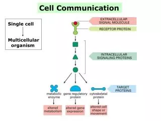
Molecular mechanisms of differentiation and programmed cell death – relation to cell pathologies Aleš Hampl. Tissues. Cells that serve various functions (supporting stucture, přijímání živin, locomotion, sensing and transfer of signals,…). Organs. Individuals (animals & plants ).

E N D
Molecular mechanisms of differentiation and programmed cell death – relation to cell pathologies Aleš Hampl
Tissues Cells that serve various functions (supporting stucture, přijímání živin, locomotion, sensing and transfer of signals,…) Organs Individuals (animals & plants) Characteristic for: complex multicellular organisms
Ontogenesis of a complex multicellular organism Zygote Multicellular embryo Dynamic multicellular organism
How does multicellular organism develop from one cell ? = one of the most critical question in biology How does this question can be answered? Ontogenesis of multicellular organism in terms of MODERN DEVELOPMENTAL BIOLOGY = Processes that drive a development of various cell types and their organisation to functional structures of living organism. Experiments that lead to understanding causative relations. Ontogenesis of multicellular organism in terms of CLASSICAL EMBRYOLOGY = Changes in numbers, localisation, and shape of cells that take place during ontogenesis. X This is only allowed by current approaches of molecular genetics and biology !
Even recent technologies of molecular biology do not make understanding regulation of development processes a trivial task !!! … because Developmental processes occuring in four dimensions: Space (x,y,z) + Time Linear information stored in genes ?
Zygote Cell division & Cell specialisation Cell division Multicellular embryo Cell division & Cell specialisation Cell division Dynamic multicellular organism
= = Proliferation Cell multiplication & Structural and functional specialisation of cells = = Differentiation & Development Cell death in predictable place and time Programmed cell death = = & Tridimensional organsiation of cells into fuctional units = = Morphogenesis
Proliferation & Differentiation Maintenance of tissue functions & Development Programmed cell death • „health“ of tissues • adaptation to • environment • repair of injured • tissues & Morphogenesis
Proliferation & Differentiation Outcomes of cell signaling & Programmed cell death & Morphogenesis
Hematopoiesis (example) Differentiation is continuos (multistep) process Increase of cell differentiation Decrease of developmental capacity - determination Stem cell Precursors (myeloid + lymphoid) Progenitors D I F F E R E N T I A T I O N Terminally differentiated cells
Once cells enter a given differentiation pathway (lineage), they don`tchange it. (for example cells of erythroid lienage never „jump“ to myeloid lineage) • Cancer cells may be the exemption from this rule • incancer, cells of a given lineage may begin to express signs • of cells of a different lineage • in cancer, it is often hard to determine from which cell lineage • cancer cells are derived • Dediferentiation – loss of differentiated phenotype • typicallyoccurs in cells explantedto in vitro • conditions (represents adaptation to culture conditions) • existence of this phenomen in vivo is still questionable • and is beiing a matter of current research (similarly to • transdiferentiation)
Zygote STABLE GENOME Genomic equivalence (= equal amount of DNA and the same nucletide sequence in all cells of a organism – cloning) Multicellular embryo X VARIABLE TRANSCRiPTOME TranscriptionRegulators Dynamic multicellular organism D I F F E R E N T I A T I O N
g a b j c i e f A C F G B D E GENERAL PRINCIPLE In every organism, all levels of organisation are typical by the dynamics of elements, from which they are made Genetic program = Genes Every level contains information that is required for building higher level. Genetic information is used stepwise based on the activity of small number of functional modules, which are hierarchically ordered with increasing level of complexity. Proteins Functional networks made of given elements develop at every level.. One part of such networks affects a lower level, the other part of such networks serves as a basis for formation of elements of higher level. Cells Tissues Organs
Factors (signals) that regulate differentiation • Soluble regulators • Hormones (glucagon, hydrocortison, tyroxin,…) • Growth factors(TGF-transforming growth factor, • FGF-fibroblast growth factor, interleukins, …) • Vitamins (D, …) • Ions (Ca++, …) • Cell-to-cell interactions (gap junctions) • Interaction of cells with extracellular matrix (colagen, laminin, …) • Polarity and shape of cells • Physical parameters of environment(temperature, tension of O2, …) These signals are processed by molecular regulatory mechanisms and networks
Universal molecular basis of regulatory mechanisms and networks • There is a relatively small number of molecular networks • These molecular networks are evolutionary highly conserved • The same molecular networks are employed in different parts • of organism to drive different processes (one molecular network interprets one • signal based on the actual molecular status of a given cell) Fibroblast growth factor - FGF
Abnormality in differentiation as a cause of disease Example Leukemic SC Hematopoietic SC Mutation Abnormality in differentiation Normal growth and differentiation Leukemic blasts Normal blood cells
Is there any use for our understanding of differentiation-regulating molecular mechanisms? • Differentiation of cells in vivo • (treatment of diseases = differentiation • therapy – cancer, …) Differentiation of cells in vitro (manipulation with stem cells = stem cell therapy)
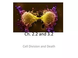
Programmed cell death A natural process for removing unwanted cells. (cells that are not needed in further development, cells with genetic abnormalities, infected cells, …)
Programmed cell death A term originally used to describe cells that die at predictable place and time during development Nearly all programmed cell death is apoptotic Apoptosis A morphological description of dying cells which contrasts with necrosis Terms are used interchangeably
APOPTOSIS NECROSIS X The stimulus of death activates a cascade of events that orchestrate the destruction of the cell The stimulus of death (e.g. ischemia) is itself a direct cause of the demise of the cell Mechanism • Chromatin condensation • Cell shrinkage • Preservation of organelles • and cell membranes • Rapid engulfment by neighboring • cells preventning inflamation • DNA fragmentation - HALLMARK • Nuclear swelling • Cell swelling • Disruption of organelles • and cell membranes • Rupture of cell and release • of cellular contents • Inflamatory response Histologic and biochemical signatures Physiologic (part of development) Aberrant (in diseases) Pathologic process
Morphological features of apoptosis • Chromatin condensation • Blebbing of cell membrane Picture in transmission electron microscope Picture in scanning electron microscope
Fragmentation of genomic DNA Internucleosomal fragments (180 bp) Electrophoretic separation of DNA in agarose gel
1972 The concept of programmed cell death first established. (Kerr, Wilie, Currie – Apoptosis: a basic biological phenomenon with wide-ranging implications in tissue kinetics. Br. J. Cancer) 2002 Nobel Prize in Physiology or Medicine „Genetic regulation of organ development and programmed cell death“ Robert Horvitz (1947) USA John Sulston (1942) UK Sydney Brenner (1927) UK
The last decade of 20ties century Key findings were obtained using Caenorhabditis elegans as a model organism(Robert Horvitz lab) What molecular mechanisms are involved in driving apoptosis? • C. elegans • organims built of exactly 1090 cells • 131 cells undergoes apoptosis during • ontogenesis • fate of all cells is well described • can be chemically mutagenized Homologs in higher metazoa ced-3 caspases Four genes that are indispensable for execution of apoptosis during ontogensis of C. elegans ced-4 Apaf-1 egl-1 Bcl-2 ced-9 BH3-only genes
Caspases – key executioners of apoptosis • Caspases = cysteinyl aspartate proteases • Proteases dependent on cysteine, they cleave after aspartic AA • Regulated primarily by posttranslational cleavage (in cells they are • present as inactive enzymes – procaspases = zymogens) • Regulated also transcriptionally (some neurodegenerative diseases are typical by • upregulated expression of caspases) • They have many substrates including themselves – this allows for • amplification of signal in cascades) • Currently 14 members of caspase family is known, 11 of which are • found in man • Not all caspases are involved in the process of programmed cell death • Caspases that participate in programmed cell death are categorized • to „initiating“ and „executing“ caspases
Proteins of BCL2 family – key regulators of apoptosis • They contain at least one region of homology with Bcl2 • (BCL2 homology region – BH) • According to their activity they divide to „antiapoptotic“ a „proapoptotic“ • BCL2 – the first identified member of this family = antiapoptotic • BAX – discovered based on its asociation with BCL2 = proapoptotic Number of AA BH4 BH3 BH1 BH2 antiapoptotic proapoptotic BH3-only
Intrinsic Extrinsic • It is initiated by binding of secreted • ligand (e.g.FasL) to „death receptor“ • that is a member of TNFR (tumor • necrosis factor rc) family (e.g. Fas) • Association with other proteins • then leads to a formation of DISC • (death-inducing signalling complex) • It is activated as a reaction on various stimuli that are generated • in cell (DNA damage, oncogene activation, oxidative stres…) • It is mediated by mitochondria, which as a reaction on stress • release proteins from their intermembrane space (cytochrome c, • SMAC, AIF, endoG…) • Cytochrome c, for example, then binds APAF1 and ATP, thus • leading to conversion of initiation procaspase-9 to active caspase-9 Two pathways initiating and executing apoptosis
Diseases & Apoptosis Abnormalitiesin apoptosisare involved in many different diseases ! Cancer Autoimunity diseases Diabetes Neurodegenerative diseases Infertility Hepatitis Sepsis Viral infections + many others Molecules reguluting apoptosis are very attractive targets for pharmacological intervention !!!
Thank you for your attention ! Questions and comments at: ahampl@med.muni.cz