Download
1 / 56
580 likes | 611 Views
Malignant Skin Tumors. Dr N. K. Kansal Associate Professor. Malignant tumor. A tumor is an abnormal mass of tissue - growth of which exceeds & is uncoordinated with that of normal tissue with capacity to metastasize to lymph nodes and other organs

E N D
Malignant Skin Tumors Dr N. K. Kansal Associate Professor
Malignant tumor • A tumor is an abnormal mass of tissue - growth of which exceeds & is uncoordinated with that of normal tissue with capacity to metastasize to lymph nodes and other organs • Deals chiefly with the malignant tumors arising from epidermal cells
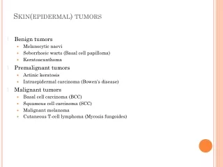
Basal cell carcinoma • Squamous cell carcinoma • Malignant melanoma
Basal cell carcinoma (BCC) • The most common cancers in humans • All BCCs - Mutations activating the Hedgehog signaling pathway • Exposure to UV light • Associated with PTCH1 gene mutation in most cases • BCCs are locally destructive but rarely metastatic • BCCs - primarily treated by surgical excision, electrodesiccation & curettage, Mohs micrographic surgery and topical agents
Epidemiology • BCC - The most common cancer in humans • Estimated - >3 million new cases occur each year in the USA • Men - affected slightly more often than are women • Tumors - More frequent in patients older than 60 years of age • Majority of BCCs- located on the head and neck
Risk factors • Risk factors for BCC - ultraviolet radiation (UVR) exposure, light hair and eye color, northern European ancestry and inability to tan • BCC is rare in dark skin - the inherent photoprotection of melanin & melanosomal dispersion
Clinical Features • Subtypes • Nodular BCC - the most common clinical subtype • Pigmented BCC - a subtype of nodular BCC that exhibits increased melanization • Superficial BCC - most commonly on the trunk • Morpheaform (sclerosing/infiltrating) BCC - an aggressive growth variant • Basosquamous carcinoma - a form of aggressive growth BCC; can be confused with squamous cell carcinoma (SCC) • Fibroepithelioma of Pinkus
Clinical Features • Presence of any nonhealinglesion Should raise the suspicion of skin cancer • BCC - usually on sun-exposed areas of the head & neck • Can occur anywhere on the body • Commonly seen features - translucency, ulceration, telangiectasias, and the presence of a rolled border • Characteristics - Differ for different clinical subtypes
Nodular BCC • The most common clinical subtype • Occurs most often on the sunexposed areas of the head & neck • Appears as a translucent papule or nodule • Usually telangiectasias and often a rolled border • Larger lesions with central necrosis - referred to by the historical term ‘rodent ulcer’
Pigmented BCC • A subtype of nodular BCC – exhibits increased melanization • Pigmented BCC - Presents as a hyperpigmented, translucent papule
Superficial BCC • Superficial BCC - most commonly on the trunk • Appears as - a well-demarcated erythematous patch • The DD nummular (discoid) dermatitis • An isolated patch of “eczema” that does not respond to treatment - raise suspicion for superficial BCC
Morpheaform BCC • An aggressive growth variant • Lesions of morpheaform BCC - have an ivorywhite appearance • May resemble a scar or a small lesion of morphea • The appearance of scar tissue [in the absence of trauma/ previous surgical procedure or the appearance of atypical-appearing scar tissue at a previously treated lesion] - alert for possibility of morpheaform BCC • The extent - often larger than the clinical appearance
Basosquamous carcinoma • A form of aggressive growth BCC • Can be confused with squamous cell carcinoma (SCC) • Histologically - Shows both basal cell and SCC differentiation in a continuous fashion
Diagnosis • Diagnosis - Accurate interpretation of the skin biopsy results • The preferred method of biopsy - shave biopsy, punch biopsy • Punch biopsy - Useful for flat lesions of morpheaform BCC & for recurrent BCC occurring in a scar • During biopsy - adequate tissue
Management • Management of BCC - guided by anatomic location & histological features • Approaches include standard surgical excision or destruction by various other physical modalities, Mohs micrographic surgery (MMS) topical chemotherapy • Best chance to cure - Through ‘adequate initial treatment’ recurrent tumors are more likely to be resistant to further treatments • May cause further local destruction
Mohs micrographic surgery • Developed in 1938 by Frederic E. Mohs, a general surgeon • A microscopically controlled surgery used to treat common types of skin cancer • During the surgery, after each removal, the tissue is examined for cancer cells • Provides informed decision for additional tissue removal • Improves prognosis - After 5 years, MMS-treated BCCs recurred in 1.4% of primary & 4% of recurrent tumors • Preferred treatment for any BCC where tissue conservation is desired
Standard surgical excision • Curettage & desiccation • Cryosurgery • Imiquimod (5% cream) • 5-Fluorouracil • Photodynamic therapy (PDT)
Squamous cell carcinoma (SCC) • SCC- second most common skin cancer, in immunocompetent after basal cell carcinoma • The most common skin cancer - in immunosuppressed organ transplantation recipients • Majority of SCC - present with early-stage disease • Prognosis - excellent in the majority of cases • Risk of developing metastasis from SCC is generallylow
Risk factors • Ultraviolet radiation (both UVB and UVA) – most important environmental risk factor for the development of SCC with a strong dose-response association • Genetic predisposition - potentiate the risk of environmental factors such as UVR • Clinical skin phenotypes - Light complexion (as in photo types I, II) • Physical & chemical carcinogens- Arsenic, used in various medications, tainted wine and unprocessed well water may stimulate skin carcinogenesis; Cutting oils - a risk of SCC development in certain industrial occupations; SCC on the scrotum of chimney sweeps - attributed to chronic exposure to ash & polycyclic aromatic hydrocarbons derived from carbon compounds (e.g. coal tar)
Immunosuppression including iatrogenic (e. g. solid organ transplantation recipients, with autoimmune or rheumatoid disease), Hematopoietic stem cell transplantation, Infection with HIV/AIDS • Viral infection – HPVs • Chronic inflammation & chronic injury of the skin – chronic ulcers (Marjolin’s), burn scars & radiation dermatitis • Chronic inflammatory disorders - discoid lupus erythematodes, mucosal & hypertrophic lichen planus, lichen sclerosus & dystrophic epidermolysis bullosa
Epidemiology • SCC incidence - increases with age; most patients =/> 60 • Higher in men than in women • Sun-sensitive individuals with red hair, blue eyes & fair complexion -higher risk than individuals with darker pigmentation • Race- Australians, exposed to very high, long-term UVR levels – more likely to develop SCC than other populations
Clinical Features • Variable & depends on the histologic subtype and location • Typically, SCCs arise on sun-exposed areas • The face, head, and neck region & the forearms & dorsum of the hands • The typical clinical finding – includes slowly enlarging, firm, skin-colored to erythematous plaques or nodules • Marked hyperkeratosis • Ulceration, exophyticor infiltrative growth patterns - seen
Verrucous SCC • Verrucous SCC - a slowly growing ulcerated plaque or an exophyticcauliflower-like slowly growing tumor • Typical locations • Oral cavity (oral florid papillomatosis) • Genitoanalregion (giant condylomaacuminatum; Buschke-Löwenstein) • Plantar skin (epitheliomacuniculatum) • Amputation stumps • Less common than other forms of invasive SCC
Diagnosis • The standard pathology report to indicate: • Histologic subtype (acantholytic, spindle cell, verrucous, or desmoplastic type) • Grade of differentiation (G1 to G4) • Maximum vertical tumor diameter in millimeters • Extent of dermal invasion (Clark level) • Presence or absence of perineural, vascular, or lymphatic invasion • Information about whether the margins are free or not
Treatment • Treatment modality for the primary lesion - major determinant for the risk of local recurrence • Ideal management - localtumor control along with maximal preservation of function and cosmesis
Surgical excision • Surgery excision - preferably microscopically controlled surgery (Mohs surgery) - primary mode of therapy • For localized lesions - cure rate of 95% • SCC - local in-transitmetastasis- may be removed by wide surgical excision or treated by irradiation of a wide field around the primary lesion • Treatment of nodal metastasis - lymph node dissection, radiation, or a combination of both
Other therapies • Topical therapeutic treatments- e.g. imiquimod, topical or intralesional 5-fluoruracil, cryotherapy & PDT – Lack of evidence for the efficacy • Radiation therapy - patient preference and other factors, e.g. problematic locations for surgery
Limited data on the efficacy of chemotherapy for metastaticSCC • Standard options in metastatic or unresectabledisease – systemic platinum-based chemotherapeutic regimens, 5- fluorouracil/capecitabine, or monotherapy/chemotherapy with methotrexate
Prognosis • Majority of SCCs- low risk • If early stage- result in a high cure rate with excellent prognosis • Prognosis for locally advanced SCC at the time of diagnosis & patients with progressive disease after first-line surgical therapy - usually poor • A poorer outcome of immunosuppressedpatients with advanced disease
Melanoma • Melanoma (Gr. melas [dark], oma[tumor]) - malignant tumor arising from melanocytic cells • Can occur anywhere where melanocytes are found • The most frequent type - cutaneous melanoma • Also at the mucosal, the uveal, or even the meningeal membrane • 10% melanomas – detected by lymph node metastases [with so-called “unknown primary”]
Epidemiology • Rising incidence worldwide - Countries with white inhabitants, with highest incidence rates in Australia (35 new cases/year/100,000) • North America (21.8 new cases/100,000) • Europe (13.5 new cases/100,000) • Median age - for melanoma diagnosis is 63 years with 15% being <45 years • Melanoma – Accounts for only 4% of all skin cancer diagnoses in the USA • Responsible for 75% of skin cancer deaths
Risk factors • History of sunburns and/or heavy sun exposure • Fitzpatrick skin phototypesI & II • Blue or green eyes, blonde or red hair, fair complexion • >100 typical nevi, or any atypical nevi • Prior personal or family history of melanoma • p16mutation
Clinical Features • Subtypes • Superficial spreading melanoma • Nodular melanoma • Lentigo maligna • Lentigo maligna melanoma • Acrallentiliginous melanoma • Desmoplastic melanoma • Mucosal melanoma
Superficial spreading melanoma • Most common subtype, accounting for approximately 70% • Most common on intermittently sunexposedareas • The lower extremity of women; the upper back of men • Irregular borders and irregular pigmentation • May present subtly as a discrete focal area of darkening • Varying shades of brown typify most melanocytic lesions • Also aspects of dark brown to black, blue-gray, red, and gray-white (which may represent regression) may be found
Nodular melanoma • Approximately 15%-30% of all melanomas • The trunk - the most common site • Remarkable for rapid evolution - often arising over several weeks to months • May lack an apparent radial growth phase • Typically appears as a uniformly dark blue-black or bluish-red raised lesion • 5% are amelanotic
Lentigo maligna & Lentigo maligna melanoma • Lentigo maligna- melanoma in situ with a prolonged radial growth phase • Eventually becomes invasive Lentigo malignamelanoma • Diagnosed most commonly in the seventh to eighth decades (uncommon before the age 40) • Most common location - the chronically sun-exposed face, on the cheeks and nose in particular • Clinical appearance - flat, slowly enlarging, brown, freckle-like macule with irregular shape & differing shades of brown and tan
Complications • Usually based on metastatic disease - symptoms associated with the affected organ • Pain (any metastases) • Convulsion (brain metastases) • Instabilities, # - (bone metastases) • Later - symptoms associated with progression of the disease & death in the palliative setting • Cutaneous changes - localized or diffuse hypo- or hyperpigmentation • Development of a melanoma-associated vitiligo [an accompanying autoimmunedisease against melanocytes] - in 4%; associated with a better prognosis