Download
1 / 62
680 likes | 1.55k Views
The Gallbladder. University of Kentucky Department of General Surgery Dustin S Campbell, MD. Grand Rounds September 28, 2011. Define the basic anatomy of the gallbladder and its relationship to surrounding structures

E N D
TheGallbladder University of Kentucky Department of General Surgery Dustin S Campbell, MD Grand Rounds September 28, 2011
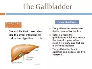
Define the basic anatomy of the gallbladder and its relationship to surrounding structures • Understand basic clinical and radiological findings in different gallbladder diseases • Discuss cost effective treatment options for choledocholithiasis • Discuss current recommendations for routine versus selective intraoperative cholangiograms Objectives
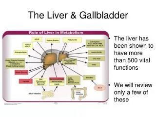
1420 gallstones identified on autopsy by pathologist Antonio Benevieni • 1630 &1637 two separate studies show the gallbladder was not an essential organ • 1743 first cholecystostomy performed by Jean-Louis Petit • 1867 John Bobbs from Indianapolis, IN opened a stone packed gallbladder and removed the gallstones leaving the gallbladder in the abdomen Gallbladder timeline De U. Evolution of cholecystectomy: A tribute to Carl August Langenbuch. Indian J Surg 2004;66:97-100.
Gallbladder timeline 1882 Carol Johann August Langenbuch performs the first cholecystectomy 1931 Pablo Luis Mirizzi performed the first cholangiography 1987 PhillipeMouret performed the first laparoscopic cholecystectomy 1992 NIH concluded laparoscopic cholecystectomy treatment of choice 1997 single port laparoscopy 2007 Natural orifice transluminal endoscopic surgery (NOTES)
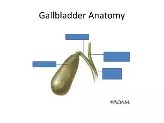
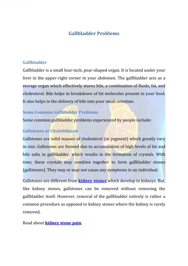
Anatomically divided into a fundus, body, infundibulum and neck • It is usually 7-10cm in length, 3-5cm in diameter and has a capacity of 30-60mL.
Blood supply to the extrahepatic biliary system orginates distally from the GDA, retroduodenaland posterosuperiorpancreatoduodenal arteries; proximally from the right hepatic & cystic arteries • These arteries supply the common bile and common hepatic ducts through branches running parallel to the duct in the 3 & 9 o’clock positions • Ischemia of the bile duct will not be readily evident at time of dissection but can result in biliary stricture or leak postoperatively
One of the leading indications for surgery in the US today, with approximately 750,000+ cholecystectomies performed every year • 10-20% of the population will develop gallstones, the incidence increases with age • Not all management is clear cut Gallstone Disease
Only about 30% of asymptomatic patients will warrant surgery during their lifetime • Landmark study by Gracie and Ransohoff followed 123 patients 15 years • 10% progressed to symptoms in 5 years • 15% by 10 years • 18% by 15 years • Overall 1-2% per year developed serious complications Asymptomatic Gallstones
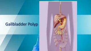
Stones greater than 2.5cm • Congenital hemolytic anemias (Sickle, hereditary spherocytosis, thalassemia) • During bariatric surgery secondary to rapid weight loss and increased lithogenicity • Polyps greater than 15mm • Transplantation ?? Cardiac only, expectant management for pancreas and/or kidney Prophylactic Cholecystectomy
Higher risk than the general population for gallstone formation • Gallstones alone are an indication for cholecystectomy • Pretransplant cholecystectomy should be considered in clinically stable patients with gallstones
Plain abdominal film: nonspecific and not useful in differentiating biliary colic and acute cholecystitis • Ultrasound: study of choice, can evaluate GB thickness, diameter of CBD, CHD, and intrahepatic ducts, pericholecystic fluid, and stones • CT scan: should not be used as an initial study for GB disease, provides similar information as US at much higher price • MRCP: expensive, very sensitive to fluid stasis and imaging CBD stones • HIDA: limited role, reproduction of symptoms with CCK injection Diagnostic Imaging
Normal Imaging hopkins-gi.org
Primary symptom—PAIN • Usually right upper quadrant, epigastric and frequently radiates to the back and right scapula • Severe enough that many patients seek medical attention with first episode • Only 50% experience pain with fatty meals • Duration of pain 1-5 hours with attacks rarely lasting longer than 24 hours Symptomatic Gallstones
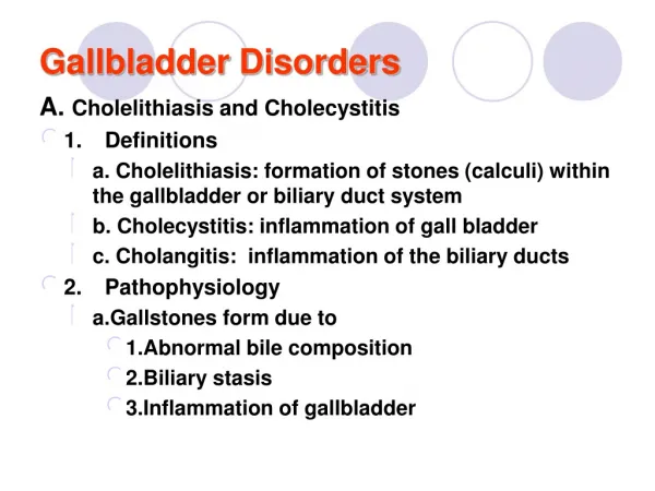
Pathogenesis • Recurrent inflammatory process involving the gallbladder • >90% patients gallstones are the causative factor • Attacks lead to scarring and nonfunctioning GB • Histopathologically CCC is characterized by an increase in subepitelial and subserosal fibrosis and mononuculear cell infiltrate Chronic CalculousCholecystitis
Presentation • Colicky pain • Chronic nausea and vomiting >60% cases • Normal physical exam • Normal lab values Chronic CalculousCholecystitis
Diagnosis • Two findings must be present—gallstones and abdominal pain consistent with biliary colic • Gallstones without symptoms do not require treatment • Management • Cholecystectomy Chronic CalculousCholecystitis
Pathophysiology • Most common complication of gallstones occurring in 20-30% of patients with symptomatic disease • Results from stone impaction at the gallbladder-cystic duct junction • In 5-20% obstruction can lead to ischemia and necrosis of the GB • Inflammatory and not an infectious process with bacterial infection appearing as a secondary event Acute CalculousCholecystitis
Presentation • Unremitting pain, may last several days and often associated with emesis, anorexia and fever • Murphy’s sign—an inspiratory arrest during deep palpation of RUQ…classic finding • Labs reveal mild leukocytosis, possible mild hyperbilirubinemia, elevated transaminases, and amylase Acute CalculousCholecystitis
Diagnosis • Ultrasound >90% sensitivity for ACC suggestive findings include GB wall thickening greater than 4mm, pericholecystic fluid and sonographic Murphy’s sign • HIDA scan may indicate and obstructed cystic duct and in the right clinical setting can have >95% sensitivity and specificity • CT scan reveals many of the same US findings, but is less sensitive and more expensive Acute CalculousCholecystitis
Treatment • Laparoscopic cholecystectomy within 24 to 72 hours of diagnosis • Early conversion to an open procedure should be considered if dissection is difficult or clear progress cannot be made • High risk patients whose medical condition(s) precludes cholecystectomy, a cholecystostomy can be performed Acute CalculousCholecystitis
Pathophysiology • Clinical cholangitis results from two factors—biliary obstruction and significant concentrations of bacteria in the bile • Most common organisms recovered—E. coli, Klebsiella, Enterococcus, Bacteroides • Biliary obstruction leads to high intrabiliary pressures resulting in bacterial organisms rapidly appearing in both blood and lymphatics Acute Cholangitis
Presentation • Wide spectrum of disease from self-limited to toxic • Charcot’s triad/Reynolds’ pentad: jaundice, fever, RUQ pain / mental status change and hypotension • Pain is usually present but much milder than acute calculouscholecystitis • Up to 33% of East Asian patients with choledocholithiasis present with toxic cholangitis Acute Cholangitis
Diagnosis • Clinical diagnosis supported by leukocytosis, hyperbilirubinemia, elevation of transaminases, US, CT, or MRCP may reveal biliary ductal dilation due to stones or mass. • Treatment • Initially supportive with antibiotics, fluids, and vasopressors if needed • Approximately 15% will not respond to antibiotics within 24 hours and require biliary decompression either percutaneous or endoscopic Acute Cholangitis
Treatment continued • In the setting of failed endoscopic and percutaneous decompression surgical CBD exploration with T-tube placement remains a life-saving procedure, but carries a significantly higher mortality • Gallstone cholangitis requires an interval cholecystectomy within 6-12 weeks as the incidence of recurrent biliary symptoms are significantly higher if the gallbladder is left in situ (6% vs 25%) Acute Cholangitis
5-10% of all patients with acute cholecystitis • Diagnosed most often in critically ill patient following trauma, burns, long term TPN, or after major nonbiliary operations such as AAA repair and cardiac bypass • Etiology unclear although stasis and ischemia are often thought to play a role • Emergency cholecystectomy if diagnosis is suspected as the incidence of gangrene, perforation and empyema exceeds 50% • Mortality upwards of 40% Acute AcalculousCholecystitis
Typical biliary colic symptoms, but no evidence of stones on US • More aggressive workup—CT scan, EGD, ERCP?, HIDA • All other studies negative and HIDA EF <35% considered abnormal • 85-94% of patients with low EF and symptoms of biliary colic treated with cholecystectomy are improved Chronic AcalculousCholecystitis
CBD stones can be classified as either primary or secondary • In the United States more than 85% are secondary • Primary duct stones typically occur with benign biliary stricture, sclerosing cholangitis, choledochal cyst disease, or sphincter of Oddi dysfunction • Primary stones require removal of the stones and a drainage procedure whereas secondary can be treated by removal of stones and cholecystectomy Choledocholithiasis
Symptoms • Anorexia • Icterus • Dark urine • H&P • Subicterus • Jaundice • History of cholangitis • History of pancreatitis Predictors of Choledocholithiasis Preoperative clinical and paraclinical predictors of choledocholithiasis
Laboratory and Imaging • Total bilirubin (>1.4 mg/dl) • Direct bilirubin (>0.3 mg/dl) • AST (>36 U/L) • ALT (>36 U/L) • CBD diameter > 6mm in the presence of stones • Observed stone on US or other imaging Predictors of Choledocholithiasis Preoperative clinical and paraclinical predictors of choledocholithiasis
Options, options and more options… • Preoperative endoscopic retrograde cholangiopancreatography ERCP • Laparoscopic cholecystectomy with intraoperative cholangiogram • Postoperative ERCP • Intraoperative ERCP • Laparoscopic common bile duct exploration • Open common bile duct exploration • Placement of a double lumen catheter Choledocholithiasis Management
One stage management of LC with IOC followed by LCBDE for positive choledocholithiasis has the lowest rate of morbidity and mortality • LCBDE does require a surgeon comfortable and facile with this technique. Sensitivity analysis reveal that when LCBDE added morbidity and mortality is 32% and 1.8% then the one stage management option will have higher morbidity and mortality rate compared to the two stage. • Decreased hospital stay with one stage technique • ERCP is preferred in patients with suppurative cholangitis, biliary sepsis or high risk surgical patients Summary
Estimated that surgeons in the United States deal with 50,000-115,000 cases of choledocholithiasis yearly • The rational allocation of scarce health care resources requires that the most cost effective approach be used to deal with such a common clinical problem • The most popular methods of detecting CBD stones include ERCP, endoscopic ultrasound, IOC, intraoperative US and MRCP Cost Analysis
The laparoscopic cholecystectomy only strategy was the least costly, but given the prevalence of CBD stones at 10% the effectiveness is only 90% • The LCBDE strategy had a cost effectiveness ratio of $5993.60, indicating that it would cost an additional $5993.60 to prevent one case of residual CBD stones • Routine preoperative ERCP is very effective at preventing residual CBD stones, approximately 333 patients would need to be managed with preop ERCP in order to avoid a single case of retained CBD stone. A cost of $299,259.35 • Preop ERCP had a better average cost effectiveness than selective postop ERCP when the prevalence of CBD stones was >0.80 Cost Analysis
Reasons for routine IOC • Screening for unsuspected BD pathology such as strictures, anomalies, and tumors • Prevention of bile duct injuries • Obtaining and maintaining proficiency • Reduction of unnecessary BDE Intraoperative Cholangiogram Evaluation of Operative Cholangiography in 2043 patients ungergoing laparoscopic cholecystectomy
Reality • Clinically significant unsuspected anomalies are very rare • Unsuspected CBD stones becoming symptomatic is 1 out of 10 and few of those will develop a complication • Injuries occur whether routine IOC is used or not…when the anatomy is clear enough to incise the cystic duct for an IOC, it is clear enough not to need an IOC Intraoperative Cholangiogram Evaluation of Operative Cholangiography in 2043 Patients undergoing Laparoscopic Cholecystectomy
Reasons against routine IOC • Prolonged operative time • Increased incidence of allergic reaction • Pancreatitis • BD injuries • Unnecessary procedures • Morbidity • Cost • Real argument—increased unnecessary morbidity and cost Intraoperative Cholangiogram Evaluation of Operative Cholangiography in 2043 Patients undergoing Laparoscopic Cholecystectomy